Explore
 Validate
 Learn
Learn Western blot
Antibody data
- Antibody Data
- Antigen structure
- References [4]
- Comments [0]
- Validations
- Western blot [1]
- Immunohistochemistry [1]
Submit
Validation data
Reference
Comment
Report error
- Product number
- AF8239 - Provider product page

- Provider
- Novus Biologicals
- Product name
- Sheep Polyclonal HB-EGF Antibody
- Antibody type
- Polyclonal
- Description
- Immunogen affinity purified. Detects mouse HB-EGF in direct ELISAs and Western Blots. In direct ELISAs, less than 30% cross-reactivity with recombinant human HB-EGF is observed and less then 2% cross-reactivity with recombinant mouse Amphiregulin is observed.
- Reactivity
- Mouse
- Host
- Sheep
- Isotype
- IgG
- Vial size
- 100 ug
- Concentration
- LYOPH
- Storage
- Use a manual defrost freezer and avoid repeated freeze-thaw cycles. 12 months from date of receipt, -20 to -70 degreesC as supplied. 1 month, 2 to 8 degreesC under sterile conditions after reconstitution. 6 months, -20 to -70 degreesC under sterile conditions after reconstitution.
Submitted references Intranasal delivery of VEGF enhances compensatory lung growth in mice.
Ablation of peri-insult generated granule cells after epilepsy onset halts disease progression.
Ablation of Newly Generated Hippocampal Granule Cells Has Disease-Modifying Effects in Epilepsy.
MicroRNA-132 enhances transition from inflammation to proliferation during wound healing.
Dao DT, Vuong JT, Anez-Bustillos L, Pan A, Mitchell PD, Fell GL, Baker MA, Bielenberg DR, Puder M
PloS one 2018;13(6):e0198700
PloS one 2018;13(6):e0198700
Ablation of peri-insult generated granule cells after epilepsy onset halts disease progression.
Hosford BE, Rowley S, Liska JP, Danzer SC
Scientific reports 2017 Dec 21;7(1):18015
Scientific reports 2017 Dec 21;7(1):18015
Ablation of Newly Generated Hippocampal Granule Cells Has Disease-Modifying Effects in Epilepsy.
Hosford BE, Liska JP, Danzer SC
The Journal of neuroscience : the official journal of the Society for Neuroscience 2016 Oct 26;36(43):11013-11023
The Journal of neuroscience : the official journal of the Society for Neuroscience 2016 Oct 26;36(43):11013-11023
MicroRNA-132 enhances transition from inflammation to proliferation during wound healing.
Li D, Wang A, Liu X, Meisgen F, Grünler J, Botusan IR, Narayanan S, Erikci E, Li X, Blomqvist L, Du L, Pivarcsi A, Sonkoly E, Chowdhury K, Catrina SB, Ståhle M, Landén NX
The Journal of clinical investigation 2015 Aug 3;125(8):3008-26
The Journal of clinical investigation 2015 Aug 3;125(8):3008-26
No comments: Submit comment
Supportive validation
- Submitted by
- Novus Biologicals (provider)
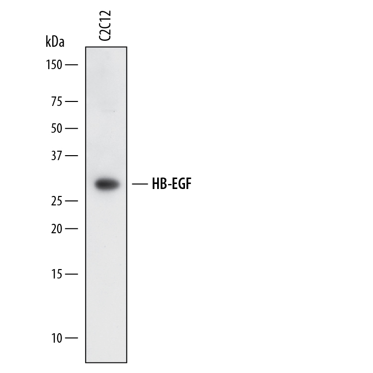
- Main image

- Experimental details
- Detection of Mouse HB-EGF by Western Blot. Western blot shows lysates of C2C12 mouse myoblast cell line. PVDF membrane was probed with 2 µg/mL of Sheep Anti-Mouse HB-EGF Antigen Affinity-purified Polyclonal Antibody (Catalog # AF8239) followed by HRP-conjugated Anti-Sheep IgG Secondary Antibody (Catalog # HAF016). A specific band was detected for HB-EGF at approximately 28 kDa (as indicated). This experiment was conducted under reducing conditions and using Immunoblot Buffer Group 1.
Supportive validation
- Submitted by
- Novus Biologicals (provider)
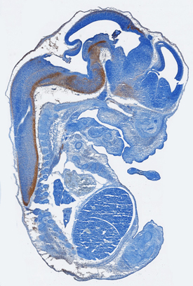
- Main image

- Experimental details
- HB-EGF in Mouse Embryo. HB-EGF was detected in immersion fixed frozen sections of mouse embryo (15 d.p.c.) using Sheep Anti-Mouse HB-EGF Antigen Affinity-purified Polyclonal Antibody (Catalog # AF8239) at 15 µg/mL overnight at 4 °C. Tissue was stained using the Anti-Sheep HRP-DAB Cell & Tissue Staining Kit (brown; Catalog # CTS019) and counterstained with hematoxylin (blue). Specific staining was localized to developing central nervous system. View our protocol for Chromogenic IHC Staining of Frozen Tissue Sections.