 Explore
 Validate
 Learn
Learn Western blot
Antibody data
- Antibody Data
- Antigen structure
- References [10]
- Comments [0]
- Validations
- Western blot [1]
- Immunohistochemistry [1]
Submit
Validation data
Reference
Comment
Report error
- Product number
- AF6295 - Provider product page

- Provider
- R&D Systems
- Product name
- Human/Mouse PRDM16/MEL1 Antibody
- Antibody type
- Polyclonal
- Description
- Antigen Affinity-purified. Detects mouse and human PRDM16 in direct ELISAs and Western blots.
- Reactivity
- Human, Mouse
- Host
- Sheep
- Conjugate
- Unconjugated
- Antigen sequence
A2A935.1- Isotype
- IgG
- Vial size
- 100 ug
- Concentration
- LYOPH
- Storage
- Use a manual defrost freezer and avoid repeated freeze-thaw cycles. 12 months from date of receipt, -20 to -70 °C as supplied. 1 month, 2 to 8 °C under sterile conditions after reconstitution. 6 months, -20 to -70 °C under sterile conditions after reconstitution.
Submitted references Cbx4 Sumoylates Prdm16 to Regulate Adipose Tissue Thermogenesis.
The Transcription Factor Prdm16 Marks a Single Retinal Ganglion Cell Subtype in the Mouse Retina.
Loss of ADAMTS5 enhances brown adipose tissue mass and promotes browning of white adipose tissue via CREB signaling.
Transcription factor Hlx controls a systematic switch from white to brown fat through Prdm16-mediated co-activation.
Fasting induces a subcutaneous-to-visceral fat switch mediated by microRNA-149-3p and suppression of PRDM16.
White-to-brown metabolic conversion of human adipocytes by JAK inhibition.
KSRP ablation enhances brown fat gene program in white adipose tissue through reduced miR-150 expression.
MicroRNA-378 controls classical brown fat expansion to counteract obesity.
MyomiR-133 regulates brown fat differentiation through Prdm16.
Brown remodeling of white adipose tissue by SirT1-dependent deacetylation of Pparγ.
Chen Q, Huang L, Pan D, Zhu LJ, Wang YX
Cell reports 2018 Mar 13;22(11):2860-2872
Cell reports 2018 Mar 13;22(11):2860-2872
The Transcription Factor Prdm16 Marks a Single Retinal Ganglion Cell Subtype in the Mouse Retina.
Groman-Lupa S, Adewumi J, Park KU, Brzezinski JA IV
Investigative ophthalmology & visual science 2017 Oct 1;58(12):5421-5433
Investigative ophthalmology & visual science 2017 Oct 1;58(12):5421-5433
Loss of ADAMTS5 enhances brown adipose tissue mass and promotes browning of white adipose tissue via CREB signaling.
Bauters D, Cobbaut M, Geys L, Van Lint J, Hemmeryckx B, Lijnen HR
Molecular metabolism 2017 Jul;6(7):715-724
Molecular metabolism 2017 Jul;6(7):715-724
Transcription factor Hlx controls a systematic switch from white to brown fat through Prdm16-mediated co-activation.
Huang L, Pan D, Chen Q, Zhu LJ, Ou J, Wabitsch M, Wang YX
Nature communications 2017 Jul 12;8(1):68
Nature communications 2017 Jul 12;8(1):68
Fasting induces a subcutaneous-to-visceral fat switch mediated by microRNA-149-3p and suppression of PRDM16.
Ding H, Zheng S, Garcia-Ruiz D, Hou D, Wei Z, Liao Z, Li L, Zhang Y, Han X, Zen K, Zhang CY, Li J, Jiang X
Nature communications 2016 May 31;7:11533
Nature communications 2016 May 31;7:11533
White-to-brown metabolic conversion of human adipocytes by JAK inhibition.
Moisan A, Lee YK, Zhang JD, Hudak CS, Meyer CA, Prummer M, Zoffmann S, Truong HH, Ebeling M, Kiialainen A, Gérard R, Xia F, Schinzel RT, Amrein KE, Cowan CA
Nature cell biology 2015 Jan;17(1):57-67
Nature cell biology 2015 Jan;17(1):57-67
KSRP ablation enhances brown fat gene program in white adipose tissue through reduced miR-150 expression.
Chou CF, Lin YY, Wang HK, Zhu X, Giovarelli M, Briata P, Gherzi R, Garvey WT, Chen CY
Diabetes 2014 Sep;63(9):2949-61
Diabetes 2014 Sep;63(9):2949-61
MicroRNA-378 controls classical brown fat expansion to counteract obesity.
Pan D, Mao C, Quattrochi B, Friedline RH, Zhu LJ, Jung DY, Kim JK, Lewis B, Wang YX
Nature communications 2014 Aug 22;5:4725
Nature communications 2014 Aug 22;5:4725
MyomiR-133 regulates brown fat differentiation through Prdm16.
Trajkovski M, Ahmed K, Esau CC, Stoffel M
Nature cell biology 2012 Dec;14(12):1330-5
Nature cell biology 2012 Dec;14(12):1330-5
Brown remodeling of white adipose tissue by SirT1-dependent deacetylation of Pparγ.
Qiang L, Wang L, Kon N, Zhao W, Lee S, Zhang Y, Rosenbaum M, Zhao Y, Gu W, Farmer SR, Accili D
Cell 2012 Aug 3;150(3):620-32
Cell 2012 Aug 3;150(3):620-32
No comments: Submit comment
Supportive validation
- Submitted by
- R&D Systems (provider)
- Main image

- Experimental details
- Detection of Human PRDM16 by Western Blot. Western blot shows lysates of K562 human chronic myelogenous leukemia cell line. PVDF Membrane was probed with 1 µg/mL of Sheep Anti-Human/Mouse PRDM16 Antigen Affinity-purified Polyclonal Antibody (Catalog # AF6295) followed by HRP-conjugated Anti-Sheep IgG Secondary Antibody (Catalog # HAF016). A specific band was detected for PRDM16 at approximately 170 kDa (as indicated). This experiment was conducted under reducing conditions and using Immunoblot Buffer Group 8.

Supportive validation
- Submitted by
- R&D Systems (provider)
- Main image
- Experimental details
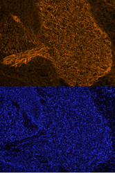
- PRDM16 in Mouse Embryo. PRDM16 was detected in immersion fixed frozen sections of mouse embryo (E13.5) using Sheep Anti-Human/Mouse PRDM16 Antigen Affinity-purified Polyclonal Antibody (Catalog # AF6295) at 10 µg/mL overnight at 4 °C. Tissue was stained using the NorthernLights™ 557-conjugated Anti-Sheep IgG Secondary Antibody (orange, upper panel; Catalog # NL010) and counterstained with DAPI (blue, lower panel). Specific staining was localized to the trigeminal ganglion. View our protocol for Fluorescent IHC Staining of Frozen Tissue Sections.
