 Explore
 Validate
 Learn
Learn Western blot
Antibody data
- Antibody Data
- Antigen structure
- References [1]
- Comments [0]
- Validations
- Western blot [1]
- Other assay [1]
Submit
Validation data
Reference
Comment
Report error
- Product number
- PA5-43407 - Provider product page

- Provider
- Invitrogen Antibodies
- Product name
- STRA6 Polyclonal Antibody
- Antibody type
- Polyclonal
- Antigen
- Synthetic peptide
- Description
- Peptide sequence: MSSQPAGNQT SPGATEDYSY GSWYIDEPQG GEELQPEGEV PSCHTSIPPG Sequence homology: Human: 100%
- Reactivity
- Human
- Host
- Rabbit
- Isotype
- IgG
- Vial size
- 100 µL
- Concentration
- 0.5 mg/mL
- Storage
- -20° C, Avoid Freeze/Thaw Cycles
Submitted references Retinol from hepatic stellate cells via STRA6 induces lipogenesis on hepatocytes during fibrosis.
Hwang I, Lee EJ, Park H, Moon D, Kim HS
Cell & bioscience 2021 Jan 6;11(1):3
Cell & bioscience 2021 Jan 6;11(1):3
No comments: Submit comment
Supportive validation
- Submitted by
- Invitrogen Antibodies (provider)
- Main image

- Experimental details
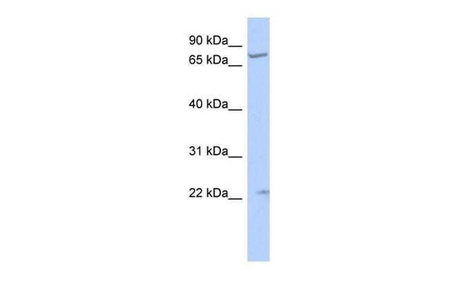
- Western blot analysis of human HepG2 cell lysate using an anti-STRA6 polyclonal antibody (Product # PA5-43407).
Supportive validation
- Submitted by
- Invitrogen Antibodies (provider)
- Main image
- Experimental details
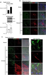
- Fig. 5 The expression of human and mouse STRA6 in vitro and in vivo. a HepG2 cells were treated with 1 muM retinol in DMSO for 1 day, and the expression levels of the human STRA6 mRNA and protein were determined. The expression level of GAPDH was detected as an endogenous control. The experiments were performed in triplicate. The quantification of western blot data was performed using Image J and unpaired Student's t-tests were performed in Prism8. * P < 0.05. b The distributions of STRA6 and alphaSMA in normal (n = 3) and TAA-induced fibrotic mouse liver tissues (n = 3). STRA6 is shown in green, and alphaSMA is shown in red. The nuclei were stained with DAPI (blue). Scale bar: 25 mum. c The distributions of STRA6 and alphaSMA in normal (n = 3) and cirrhotic human liver tissues (n = 4). STRA6 is shown in green, and alphaSMA is shown in red. The nuclei were stained with DAPI (blue). Four samples in the cirrhosis group were analyzed independently, and representative data are shown in each panel. Scale bar of lower magnification: 25 mum; scale bar of higher magnification: 5 mum. d The accumulation of triglyceride in HepG2 cells treated with or without 1 muM retinol in DMSO for 24 h following transfection with a siRNA targeting human STRA6. Retinol autofluorescence is shown in blue, and triglyceride was stained using BODIPY (red). Retinol or triglyceride-positive area % per 0.03 mm 2 were quantified five images using Image J program. Reproducible result from three independent exp
