 Explore
 Validate
 Learn
Learn Western blot
 Immunohistochemistry
Antibody data
- Antibody Data
- Antigen structure
- References [4]
- Comments [0]
- Validations
- Western blot [2]
- Immunohistochemistry [5]
Submit
Validation data
Reference
Comment
Report error
- Product number
- HPA000646 - Provider product page

- Provider
- Atlas Antibodies
- Proper citation
- Atlas Antibodies Cat#HPA000646, RRID:AB_1078935
- Product name
- Anti-LGALS1
- Antibody type
- Polyclonal
- Reactivity
- Human, Mouse, Rat
- Host
- Rabbit
- Conjugate
- Unconjugated
- Antigen sequence
EVAPDAKSFVLNLGKDSNNLCLHFNPRFNAHGDAN
TIVCNSKDGGAWGTEQREAVFPFQPGSVAEVCITF
DQANLTVKLPDGYEFKFPNRLNLEAINYMAADGDF
KIKCV- Isotype
- IgG
- Vial size
- 100 µl
- Storage
- Store at +4°C for short term storage. Long time storage is recommended at -20°C.
Submitted references Systems-level effects of ectopic galectin-7 reconstitution in cervical cancer and its microenvironment.
Immunocytochemical characterisation of olfactory ensheathing cells of zebrafish.
The protein expression of TRP-1 and galectin-1 in cutaneous malignant melanomas.
From gene expression analysis to tissue microarrays: a rational approach to identify therapeutic and diagnostic targets in lymphoid malignancies.
Higareda-Almaraz JC, Ruiz-Moreno JS, Klimentova J, Barbieri D, Salvador-Gallego R, Ly R, Valtierra-Gutierrez IA, Dinsart C, Rabinovich GA, Stulik J, Rösl F, Rincon-Orozco B
BMC cancer 2016 Aug 24;16(1):680
BMC cancer 2016 Aug 24;16(1):680
Immunocytochemical characterisation of olfactory ensheathing cells of zebrafish.
Lazzari M, Bettini S, Franceschini V
Journal of anatomy 2014 Feb;224(2):192-206
Journal of anatomy 2014 Feb;224(2):192-206
The protein expression of TRP-1 and galectin-1 in cutaneous malignant melanomas.
Bolander A, Agnarsdóttir M, Strömberg S, Ponten F, Hesselius P, Uhlen M, Bergqvist M
Cancer genomics & proteomics 2008 Nov-Dec;5(6):293-300
Cancer genomics & proteomics 2008 Nov-Dec;5(6):293-300
From gene expression analysis to tissue microarrays: a rational approach to identify therapeutic and diagnostic targets in lymphoid malignancies.
Ek S, Andréasson U, Hober S, Kampf C, Pontén F, Uhlén M, Merz H, Borrebaeck CA
Molecular & cellular proteomics : MCP 2006 Jun;5(6):1072-81
Molecular & cellular proteomics : MCP 2006 Jun;5(6):1072-81
No comments: Submit comment
Enhanced validation
Enhanced validation
- Submitted by
- Atlas Antibodies (provider)
- Enhanced method
- Orthogonal validation
- Main image
- Experimental details
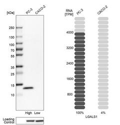
- Western blot analysis in human cell lines PC-3 and Caco-2 using Anti-LGALS1 antibody. Corresponding LGALS1 RNA-seq data are presented for the same cell lines. Loading control: Anti-HSP90B1.

Enhanced validation
- Submitted by
- Atlas Antibodies (provider)
- Enhanced method
- Independent antibody validation
- Main image

- Experimental details
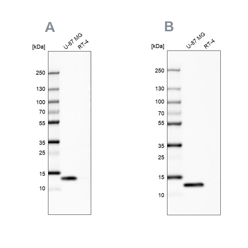
- Western blot analysis using Anti-LGALS1 antibody HPA000646 (A) shows similar pattern to independent antibody HPA000687 (B).
Supportive validation
- Submitted by
- Atlas Antibodies (provider)
- Main image
- Experimental details
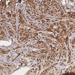
- Immunohistochemical staining of human smooth muscle shows cytoplasmic positivity in smooth muscle cells.
- Sample type
- HUMAN

- Submitted by
- Atlas Antibodies (provider)
- Main image
- Experimental details
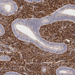
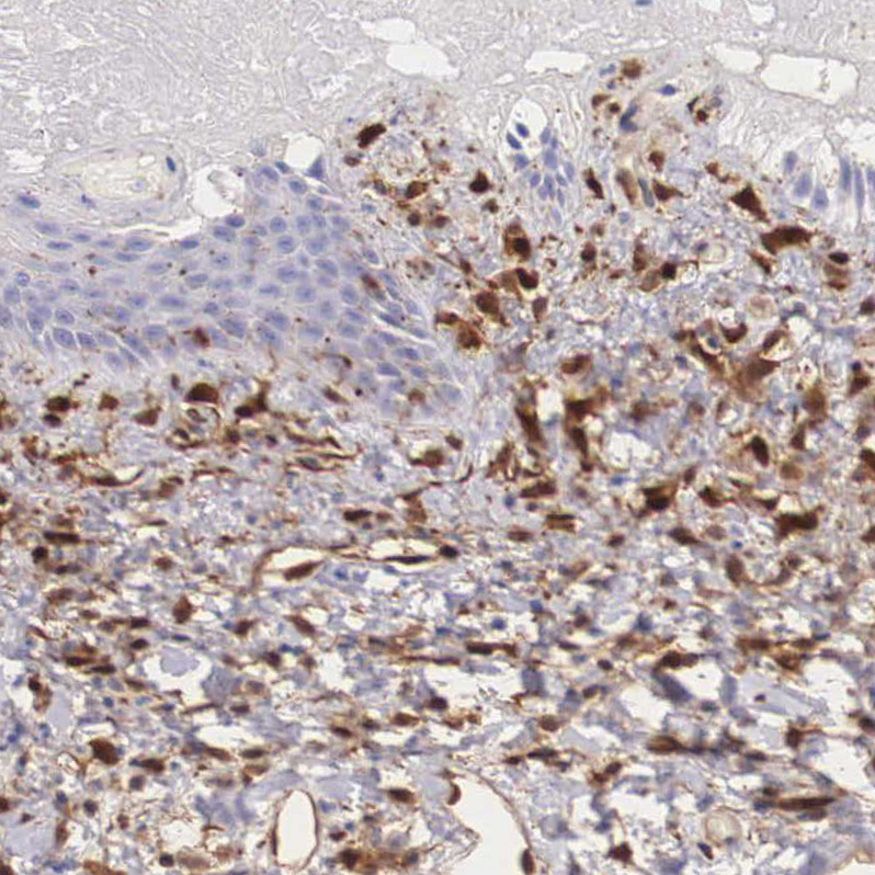
- Immunohistochemical staining of human endometrium shows strong cytoplasmic positivity in cells in endometrial stroma.
- Sample type
- HUMAN

- Submitted by
- Atlas Antibodies (provider)
- Main image
- Experimental details
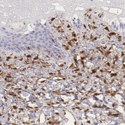
- Immunohistochemical staining of human skin shows moderate cytoplasmic positivity in dermal cells.
- Sample type
- HUMAN
- Submitted by
- Atlas Antibodies (provider)
- Main image
- Experimental details
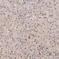
- Immunohistochemical staining of human liver shows moderate cytoplasmic positivity in a subset of hepatocytes.
- Sample type
- HUMAN

- Submitted by
- Atlas Antibodies (provider)
- Main image
- Experimental details
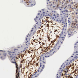
- Immunohistochemical staining of human placenta shows moderate cytoplasmic positivity in decidual cells.
- Sample type
- HUMAN
