 Explore
 Validate
 Learn
Learn Western blot
Antibody data
- Antibody Data
- Antigen structure
- References [0]
- Comments [0]
- Validations
- Western blot [3]
- Immunocytochemistry [2]
- Immunohistochemistry [5]
Submit
Validation data
Reference
Comment
Report error
- Product number
- UM800035 - Provider product page

- Provider
- OriGene
- Proper citation
- OriGene Cat#UM800035, RRID:AB_2629147
- Product name
- VSNL1 mouse monoclonal antibody,clone UMAB116
- Antibody type
- Monoclonal
- Description
- VSNL1 mouse monoclonal antibody,clone UMAB116
- Host
- Mouse
- Conjugate
- Unconjugated
- Epitope
- VSNL1
- Isotype
- IgG
- Antibody clone number
- UMAB116
- Vial size
- 100 µl
- Concentration
- 1.00mg/ml
No comments: Submit comment
Supportive validation
- Submitted by
- OriGene (provider)
- Main image

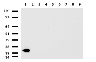
- Experimental details
- Western blot of cell lysates (35ug) from 9 different cell lines (1: HepG2, 2: HeLa, 3: SV-T2, 4: A549. 5: COS7, 6: Jurkat, 7: MDCK, 8: PC-12, 9: MCF7).
- Validation comment
- WB
- Submitted by
- OriGene (provider)
- Main image

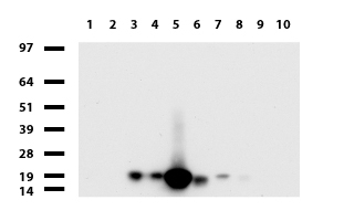
- Experimental details
- Western blot of human tissue lysates (15ug) from 10 different tissues (1: Testis, 2: Omentum, 3: Uterus, 4: Breast, 5: Brain, 6: Liver, 7: Ovary, 8: Colon, 9: Spleen, 10: Thyroid). Diluation: 1:500.
- Validation comment
- WB
- Submitted by
- OriGene (provider)
- Main image

- Experimental details
- Western blot of mouse tissue lysates (20ug) from Brian. Primary antibody diluation: 1:500. Secondary antibody dilution: Mouse TrueBlot? Ultra (1:1000).
- Validation comment
- WB

Supportive validation
- Submitted by
- OriGene (provider)
- Main image
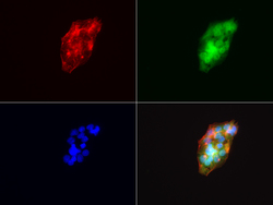
- Experimental details
- Immunofluorescent staining of HepG2 cells using anti-VSNL1 mouse monoclonal antibody (UM800035, green, 1:100). Actin filaments were labeled with Alexa Fluor? 594 Phalloidin (red), and nuclear with DAPI (blue).
- Validation comment
- IF

- Submitted by
- OriGene (provider)
- Main image
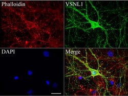
- Experimental details
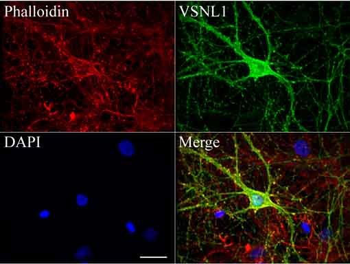
- Confocal immunofluoresce image of primary rat neurons labeled with anti-VSNL1 mouse monoclonal antibody (UM800035, green, 1:100). Actin filaments were labeled with TRICT-Phalloidin (red), and nuclear with DAPI (blue). Scale bar, 20?m.
- Validation comment
- IF
Supportive validation
- Submitted by
- OriGene (provider)
- Main image
- Experimental details
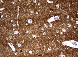
- Immunohistochemical staining of paraffin-embedded Human adult brain tissue using anti-VSNL1 mouse monoclonal antibody. (UM800035; heat-induced epitope retrieval by 10mM citric buffer, pH6.0, 120C for 3min)
- Submitted by
- OriGene (provider)
- Main image

- Experimental details
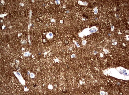
- Immunohistochemical staining of paraffin-embedded Human embryonic brain cortex tissue using anti-VSNL1 mouse monoclonal antibody. (UM800035; heat-induced epitope retrieval by 10mM citric buffer, pH6.0, 120C for 3min)

- Submitted by
- OriGene (provider)
- Main image
- Experimental details
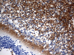
- Immunohistochemical staining of paraffin-embedded Human embryonic cerebellum using anti-VSNL1 mouse monoclonal antibody. (UM800035; heat-induced epitope retrieval by 10mM citric buffer, pH6.0, 120C for 3min)
- Validation comment
- IHC

- Submitted by
- OriGene (provider)
- Main image
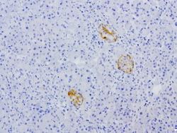
- Experimental details
- Immunohistochemical staining of paraffin-embedded human pancreas using anti-VSNL1 clone UMAB116 mouse monoclonal antibody (UM800035) at 1:200 with Polink2 Broad HRP DAB detection kit; heat-induced epitope retrieval with GBI Citrate pH6.0 HIER buffer using
- Validation comment
- IHC
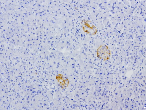
- Submitted by
- OriGene (provider)
- Main image
- Experimental details
- Immunohistochemical staining of paraffin-embedded human brain using anti-VSNL1 clone UMAB116 mouse monoclonal antibody (UM800035) at 1:200 with Polink2 Broad HRP DAB detection kit; heat-induced epitope retrieval with GBI Citrate pH6.0 HIER buffer using pr
- Validation comment
- IHC
