Explore
 Validate
 Learn
LearnPA5-77092
antibody from Invitrogen Antibodies
Targeting: VSNL1
HLP3, HPCAL3, HUVISL1, VILIP, VILIP-1
 Western blot
Antibody data
- Antibody Data
- Antigen structure
- References [1]
- Comments [0]
- Validations
- Western blot [1]
- Other assay [4]
Submit
Validation data
Reference
Comment
Report error
- Product number
- PA5-77092 - Provider product page

- Provider
- Invitrogen Antibodies
- Product name
- VSNL1 Polyclonal Antibody
- Antibody type
- Polyclonal
- Antigen
- Recombinant full-length protein
- Reactivity
- Human, Mouse, Rat
- Host
- Rabbit
- Isotype
- IgG
- Vial size
- 100 µL
- Concentration
- 1 mg/mL
- Storage
- Store at 4°C short term. For long term storage, store at -20°C, avoiding freeze/thaw cycles.
Submitted references Cdc25A inhibits autophagy-mediated ferroptosis by upregulating ErbB2 through PKM2 dephosphorylation in cervical cancer cells.
Wang C, Zeng J, Li LJ, Xue M, He SL
Cell death & disease 2021 Nov 6;12(11):1055
Cell death & disease 2021 Nov 6;12(11):1055
No comments: Submit comment
Supportive validation
- Submitted by
- Invitrogen Antibodies (provider)
- Main image
- Experimental details
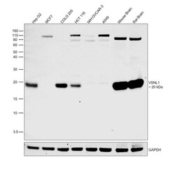
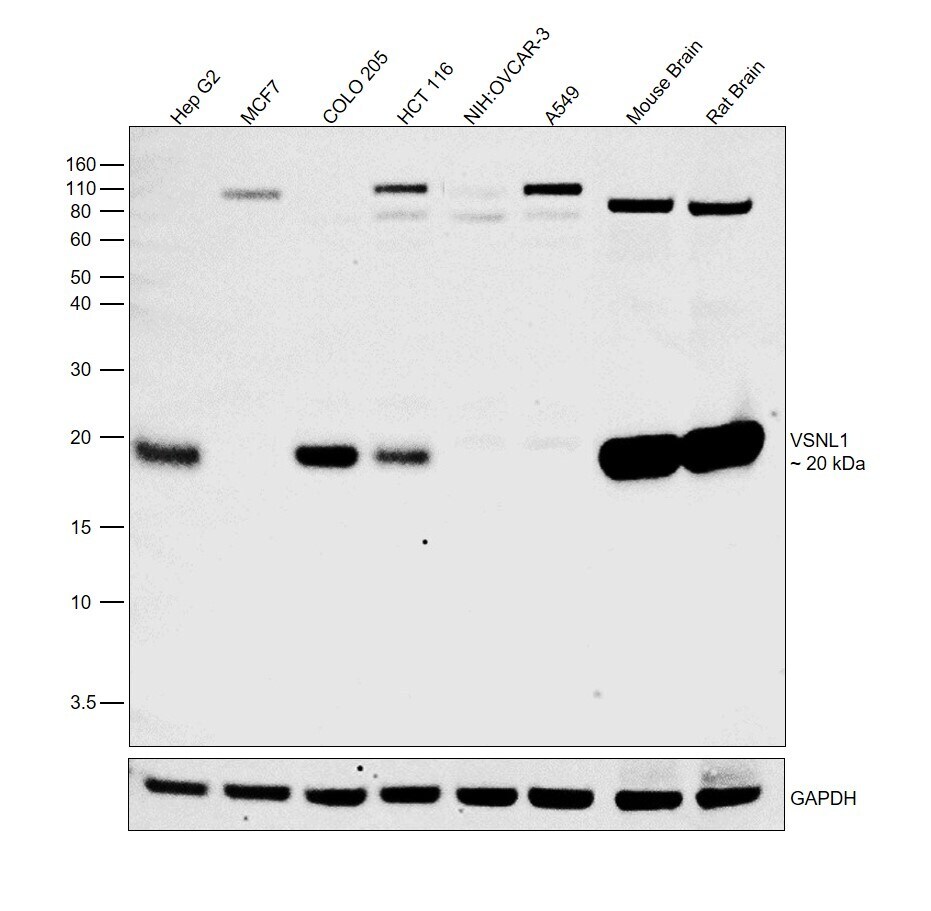
- Western blot was performed using Anti-VSNL1 Polyclonal Antibody (Product # PA5-77092) and a 20kDa band corresponding to VSNL1 was observed. Whole cell extracts (30 µg lysate) of Hep G2 (Lane 1), MCF7 (Lane 2), COLO 205 (Lane 3), HCT 116 (Lane 4), NIH:OVCAR-3 (Lane 5), A549 (Lane 6) and tissue extracts of Mouse Brain (Lane 7), Rat Brain (Lane 8) were electrophoresed using NuPAGE™ 10% Bis-Tris Protein Gel (Product # NP0302BOX). Resolved proteins were then transferred onto a Nitrocellulose membrane (Product # IB23001) by iBlot® 2 Dry Blotting System (Product # IB21001). The blot was probed with the primary antibody (1:1000) and detected by chemiluminescence with Goat anti-Rabbit IgG (H+L) Superclonal™ Recombinant Secondary Antibody, HRP (Product # A27036,1:4000) using the iBright FL 1000 (Product # A32752). Chemiluminescent detection was performed using Novex® ECL Chemiluminescent Substrate Reagent Kit (Product # WP20005). Relative expression of VSNL1 was observed between COLO 205, HCT 116 and NIH:OVCAR-3, A549 as reported in the literature (DOI: 10.1371/journal.pone.0001698). Relative expression was also observed between Hep G2 and MCF7, a cell line that expresses VSNL1 at very low levels.
Supportive validation
- Submitted by
- Invitrogen Antibodies (provider)
- Main image
- Experimental details
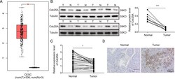
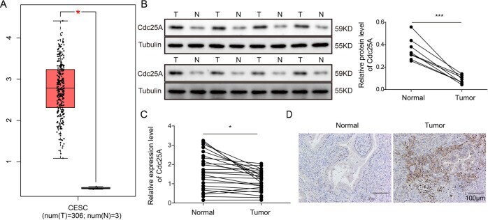
- Fig. 1 Cdc25A was elevated in human cervical cancer tissues. A Cdc25A levels in cervical cancer tissues and normal cervical tissues from the TCGA dataset. B Cdc25A protein levels in human cervical cancer tissues and adjacent non-tumour tissues from eight cervical cancer patients. C Cdc25A mRNA levels in human cervical cancer tissues and adjacent non-tumour tissues from cervical cancer patients ( N = 24). D IHC analysis of Cdc25A levels in human cervical cancer tissues and adjacent non-tumour tissues from cervical cancer patients. Each experiment was repeated three times. * P < 0.05; *** P < 0.001.
- Submitted by
- Invitrogen Antibodies (provider)
- Main image
- Experimental details
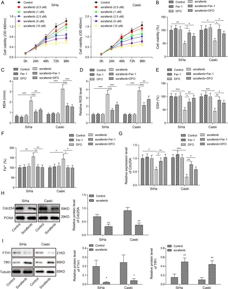
- Fig. 2 Cdc25A was reduced during sorafenib-induced ferroptosis of human cervical cancer cells. A The MTT assay to analyse cell viability following sorafenib treatment with different concentrations. B The MTT assay to analyse cell viability upon various drug treatments. C Cellular MDA levels after drug treatment. D Cellular ROS levels after 48 h of drug treatment. E Cellular GSH levels after 48 h of drug treatment. F Cellular iron levels after 48 h of drug treatment. G and H Cdc25A mRNA ( G ) and protien levels ( H ) in cervical cancer cells following 48 h of sorafenib treatment. I Protein levels of FTH1 and TfR1 in cervical cancer cells following 48 h of sorafenib treatment. Each experiment was repeated three times. * P < 0.05; ** P < 0.01; *** P < 0.001.
- Submitted by
- Invitrogen Antibodies (provider)
- Main image
- Experimental details
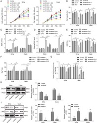
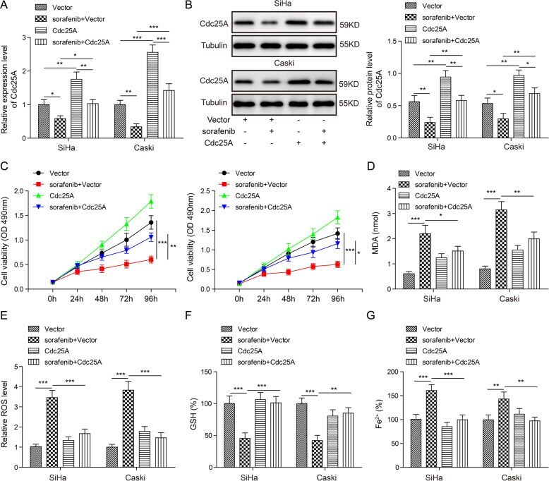
- Fig. 3 Overexpression of Cdc25A suppressed sorafenib-induced ferroptosis. A Relative Cdc25A mRNA levels in transfected cells with or without sorafenib treatment. B Relative Cdc25A protein levels in transfected cells with or without sorafenib treatment. C The MTT assay to analyse the viability of transfected cells following sorafenib treatment. D Cellular MDA levels in transfected cells after sorafenib treatment. E Cellular ROS levels in transfected cells after sorafenib treatment. F Cellular GSH levels in transfected cells after sorafenib treatment. G Cellular iron levels in transfected cells after sorafenib treatment. Each experiment was repeated three times. * P < 0.05; ** P < 0.01; *** P < 0.001.
- Submitted by
- Invitrogen Antibodies (provider)
- Main image
- Experimental details
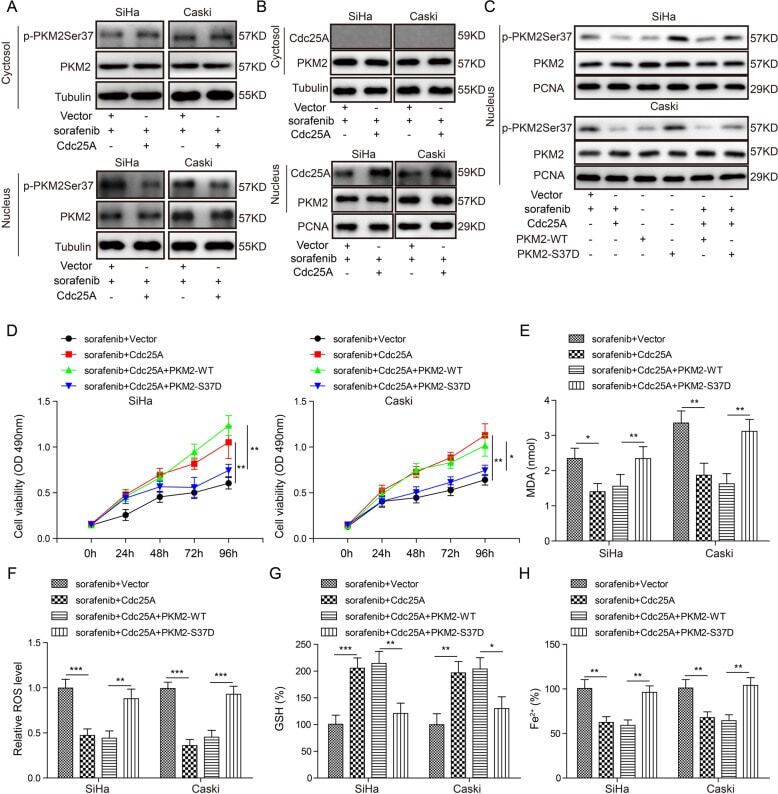
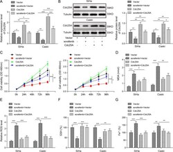
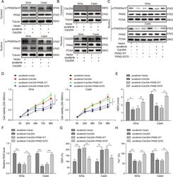
- Fig. 4 Cdc25A dephosphorylated PKM2 to suppress sorafenib-induced ferroptosis. A Relative p-PKM2 Ser37 levels in the nucleus and cytosol of transfected cells following sorafenib treatment. B Co-IP was used to analyse the interaction of Cdc25A and PKM2 in the nucleus and cytosol. C Relative p-PKM2 Ser37 levels in transfected cells with or without sorafenib treatment. D The MTT assay to analyse the viability of transfected cells following sorafenib treatment. E Cellular MDA levels in transfected cells after sorafenib treatment. F Cellular ROS levels in transfected cells after sorafenib treatment. G Cellular GSH levels in transfected cells after sorafenib treatment. H Cellular iron levels in transfected cells after sorafenib treatment. Each experiment was repeated three times. * P < 0.05; ** P < 0.01; *** P < 0.001.