 Explore
 Validate
 Learn
LearnMA5-29385
antibody from Invitrogen Antibodies
Targeting: LAMP1
CD107a
 Western blot
 ELISA Immunocytochemistry
 Immunoprecipitation Immunohistochemistry Flow cytometry Other assay
Antibody data
- Antibody Data
- Antigen structure
- References [1]
- Comments [0]
- Validations
- Western blot [4]
- Immunocytochemistry [3]
- Immunohistochemistry [2]
- Flow cytometry [1]
- Other assay [2]
Submit
Validation data
Reference
Comment
Report error
- Product number
- MA5-29385 - Provider product page

- Provider
- Invitrogen Antibodies
- Product name
- LAMP1 Recombinant Rabbit Monoclonal Antibody (107)
- Antibody type
- Monoclonal
- Antigen
- Recombinant full-length protein
- Reactivity
- Human, Rat
- Host
- Rabbit
- Isotype
- IgG
- Antibody clone number
- 107
- Vial size
- 100 µL
- Concentration
- 1 mg/mL
- Storage
- Store at 4°C short term. For long term storage, store at -20°C, avoiding freeze/thaw cycles.
Submitted references Application of meso-CF(3)-Fluorophore BODIPY with Phenyl and Pyrazolyl Substituents for Lifetime Visualization of Lysosomes.
Trukhan IS, Tomilin DN, Dremina NN, Sobenina LN, Shurygin MG, Petrushenko KB, Petrushenko IK, Trofimov BA, Shurygina IA
Molecules (Basel, Switzerland) 2022 Aug 7;27(15)
Molecules (Basel, Switzerland) 2022 Aug 7;27(15)
No comments: Submit comment
Supportive validation
- Submitted by
- Invitrogen Antibodies (provider)
- Main image

- Experimental details
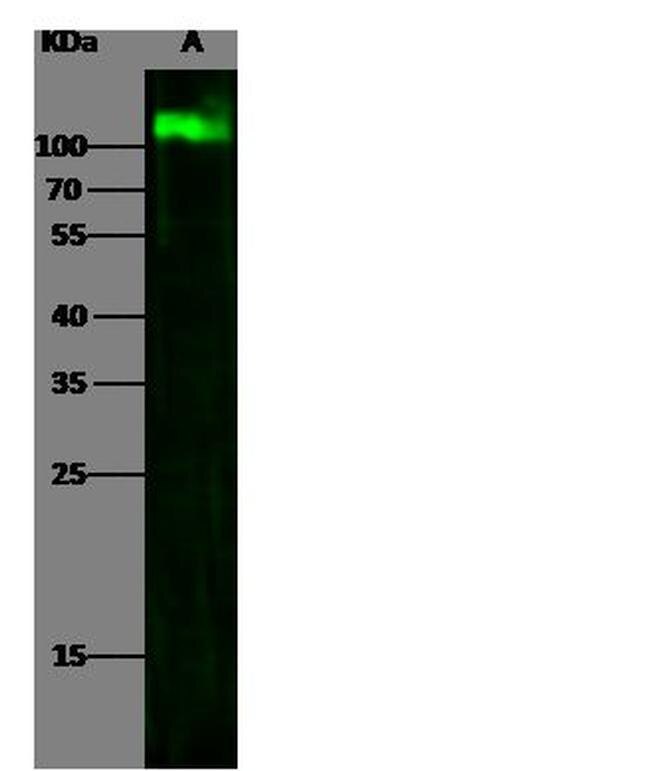
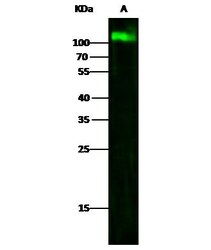
- Western blot analysis of LAMP1 in Lane A: Jurkat Whole Cell Lysate (30 µg). Samples were probed using a LAMP1 Monoclonal Antibody (Product # MA5-29385) at a 1:500 dilution, followed by a Goat Anti-Rabbit IgG (H+L), Dylight 800 Secondary Antibody at a 1:10000 dilution. Western blot was performed under reducing conditions. Predicted band size:120 kDa. Observed band size:120 kDa.
- Submitted by
- Invitrogen Antibodies (provider)
- Main image
- Experimental details
- Western Blot using LAMP1 Recombinant Rabbit Monoclonal Antibody (107) (Product # MA5-29385) at 1:500 dilution. Lane A: Jurkat Whole Cell Lysate. Lysates/proteins at 30 μg per lane. Secondary Goat Anti-Rabbit IgG H&L (DyLight™ 800) at 1:10,000 dilution. Developed using the Odyssey technique. Performed under reducing conditions. Predicted band size: 120 kDa. Observed band size: 120 kDa.

- Submitted by
- Invitrogen Antibodies (provider)
- Main image
- Experimental details
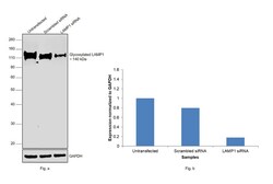
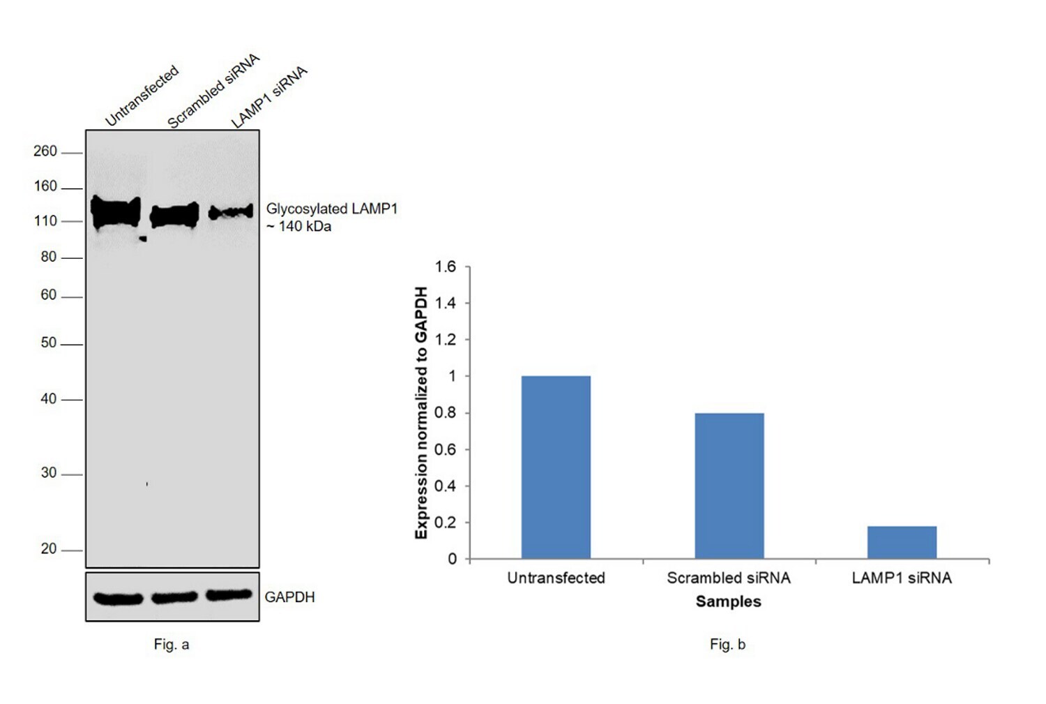
- Knockdown of LAMP1 (Lysosome-associated membrane glycoprotein 1) was achieved by transfecting Hep G2 with LAMP1 specific siRNAs (Silencer® select Product # s8081, s8082). Western blot analysis (Fig. a) was performed using whole cell extracts from the LAMP1 knockdown cells (lane 3), non-targeting scrambled siRNA transfected cells (lane 2) and untransfected cells (lane 1). The blot was probed with LAMP1 Recombinant Rabbit Monoclonal Antibody (107) (Product # MA5-29385, 1:1000 dilution) and Goat anti-Rabbit IgG (H+L) Superclonal™ Recombinant Secondary Antibody, HRP (Product # A27036, 1:10000 dilution). Densitometric analysis of this western blot is shown in histogram (Fig. b). Decrease in signal upon siRNA mediated knock down confirms that antibody is specific to Lysosome-associated membrane glycoprotein 1.
- Submitted by
- Invitrogen Antibodies (provider)
- Main image
- Experimental details
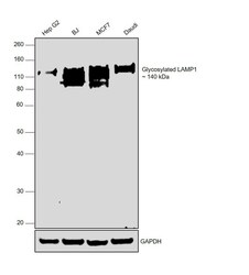
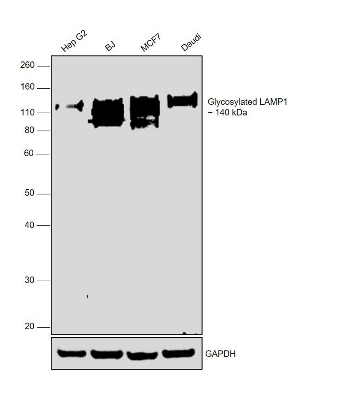
- Western blot was performed using Anti-LAMP1 Recombinant Rabbit Monoclonal Antibody (107) (Product # MA5-29385) and a 140kDa band corresponding to CD107a/LAMP1 (Lysosome-associated membrane glycoprotein 1) was observed across cell lines tested. Whole cell extracts (40 µg lysate) of Hep G2 (Lane 1), BJ (Lane 2), MCF7 (Lane 3) and Daudi (Lane 4) were electrophoresed using NuPAGE™ 4-12% Bis-Tris Protein Gel (Product # NP0321BOX). Resolved proteins were then transferred onto a Nitrocellulose membrane (Product # IB23001) by iBlot® 2 Dry Blotting System (Product # IB21001). The blot was probed with the primary antibody (1:1500 dilution) and detected by chemiluminescence with Goat anti-Rabbit IgG (H+L) Superclonal™ Recombinant Secondary Antibody, HRP (Product # A27036,1:10000 dilution) using the iBright FL 1000 (Product # A32752). Chemiluminescent detection was performed using Novex® ECL Chemiluminescent Substrate Reagent Kit (Product # WP20005). LAMP1 is a heavily glycosylated protein and is reported to show bands above ~110kDa
Supportive validation
- Submitted by
- Invitrogen Antibodies (provider)
- Main image
- Experimental details
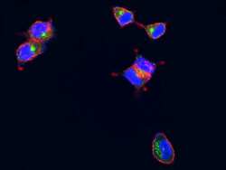
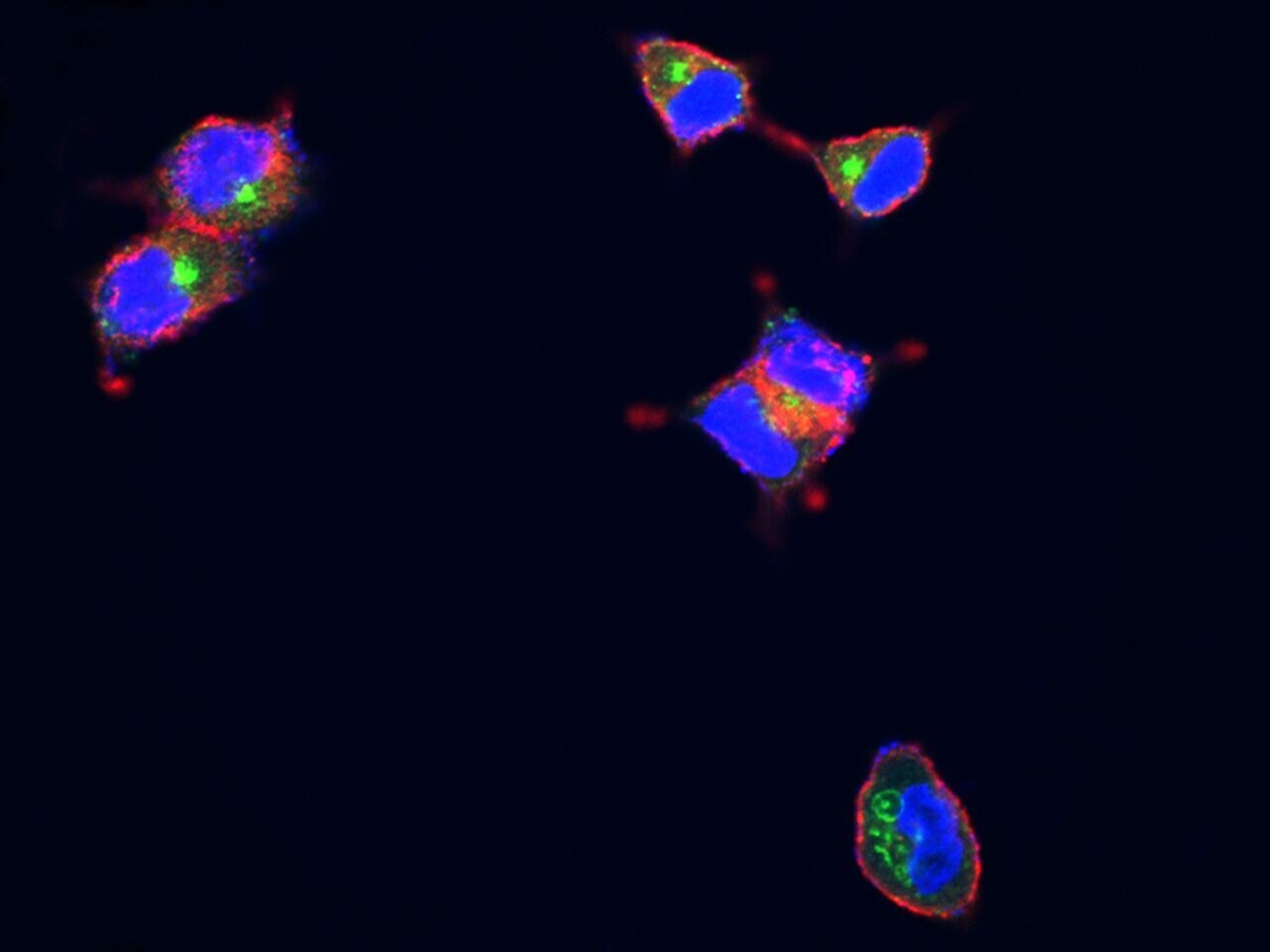
- Confocal immunofluorescence analysis of Human LAMP1 in MCF7 cells. Cells were fixed with 4% PFA, permeabilzed with 1% Triton X-100 in PBS, blocked with 10% serum, and incubated with LAMP1 Recombinant Rabbit Monoclonal Antibody (107) (Product # MA5-29385, 1:300). Then cells were stained with the Alexa Fluor® 488-conjugated Goat Anti-rabbit IgG secondary antibody, counterstained with Alexa Fluor® 546-conjugated phallotoxins (red) and DAPI (blue). Positive staining was localized to lysosome membrane.

- Submitted by
- Invitrogen Antibodies (provider)
- Main image

- Experimental details
- Confocal immunofluorescence analysis of Human LAMP1 in MCF7 cells. Cells were fixed with 4% PFA, permeabilzed with 1% Triton X-100 in PBS, blocked with 10% serum, and incubated with LAMP1 Recombinant Rabbit Monoclonal Antibody (107) (Product # MA5-29385, 1:300). Then cells were stained with the Alexa Fluor® 488-conjugated Goat Anti-rabbit IgG secondary antibody, counterstained with Alexa Fluor® 546-conjugated phallotoxins (red) and DAPI (blue). Positive staining was localized to lysosome membrane.
- Submitted by
- Invitrogen Antibodies (provider)
- Main image
- Experimental details
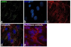
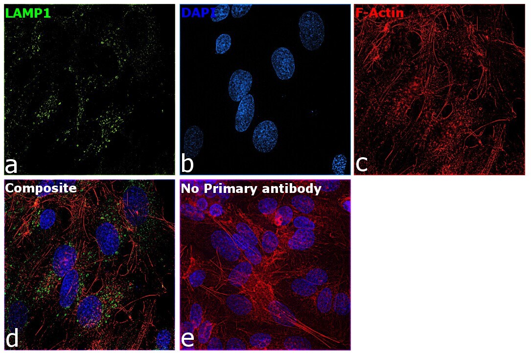
- Immunofluorescence analysis of LAMP1 (Lysosome-associated membrane glycoprotein 1) was performed using 80% confluent log phase Hep G2 cells. The cells were fixed with 4% paraformaldehyde for 10 minutes, permeabilized with 0.1% Triton™ X-100 for 10 minutes, and blocked with 2% BSA for 45 minutes at room temperature. The cells were labeled with LAMP1 Recombinant Rabbit Monoclonal Antibody (107) (Product # MA5-29385) at 1:200 in 0.1% BSA, incubated at 4 degree celsius overnight and then labeled with Donkey anti-Rabbit IgG (H+L) Highly Cross-Adsorbed Secondary Antibody, Alexa Fluor Plus 488 (Product # A32790), (1:2500 dilution), for 45 minutes at room temperature (Panel a: Green). Nuclei (Panel b:Blue) were stained with ProLong™ Diamond Antifade Mountant with DAPI (Product # P36962). F-actin (Panel c: Red) was stained with Rhodamine Phalloidin (Product # R415, 1:300). Panel d represents the merged image showing endosome and lysosome-like staining for LAMP1. Panel e represents control Hep G2 cells with no primary antibody to assess background. The images were captured at 60X magnification.
Supportive validation
- Submitted by
- Invitrogen Antibodies (provider)
- Main image
- Experimental details
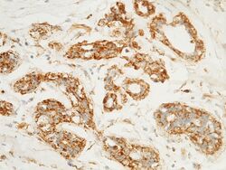
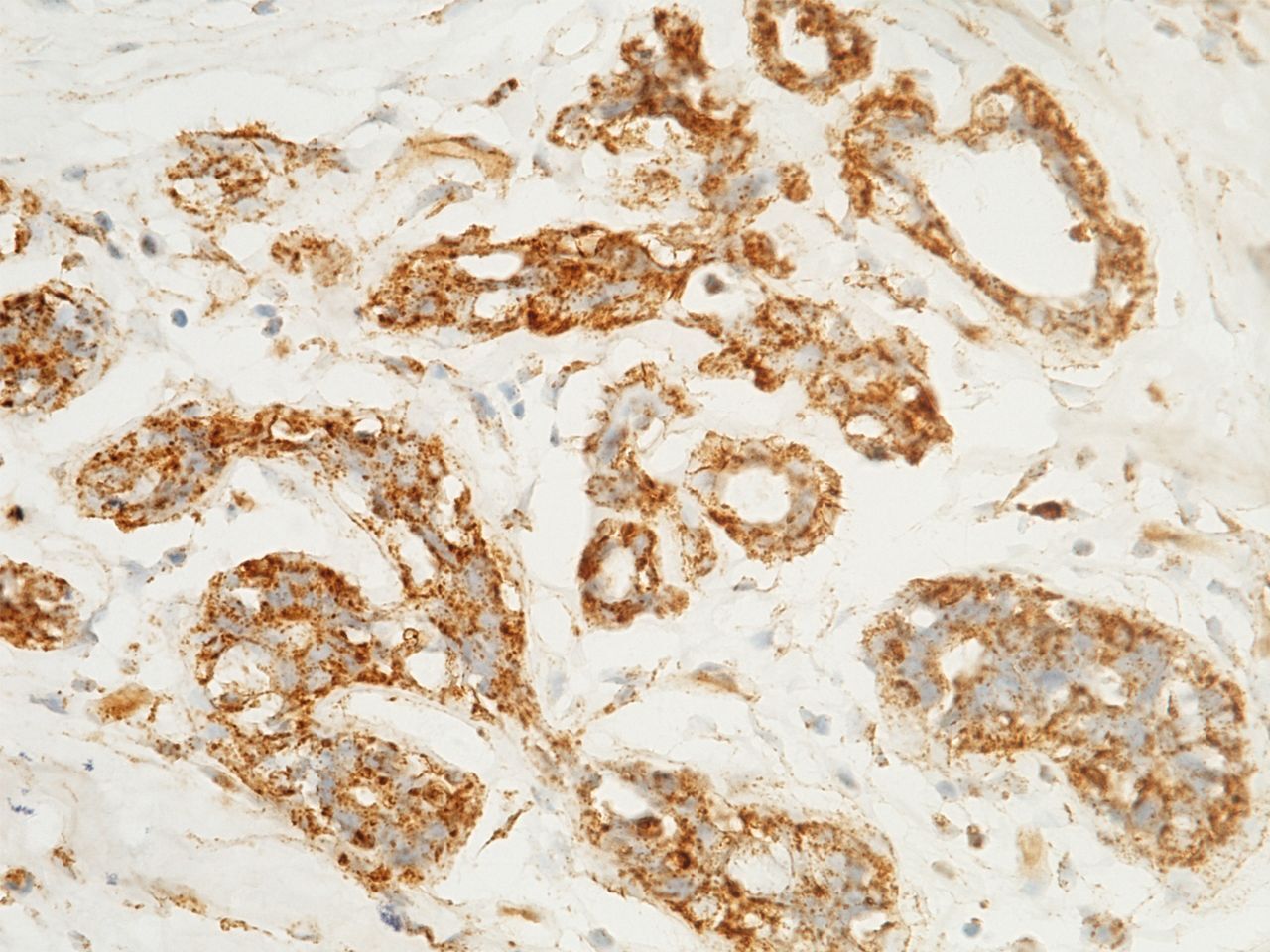
- Immunohistochemical staining of human LAMP1 in human breast carcinoma with LAMP1 Recombinant Rabbit Monoclonal Antibody (107) (Product # MA5-29385, 1:1,000, formalin-fixed paraffin embedded sections).
- Submitted by
- Invitrogen Antibodies (provider)
- Main image
- Experimental details
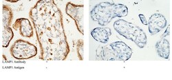
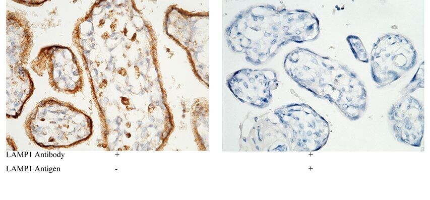
- Immunohistochemical staining of human LAMP1 in human placenta with LAMP1 Recombinant Rabbit Monoclonal Antibody (107) (Product # MA5-29385, 1:1,000, formalin-fixed paraffin embedded sections). Left panel: tissue incubated with primary antibody. Right panel: tissue incubated with mixture of primary antibody and antigen (recombinant protein).
Supportive validation
- Submitted by
- Invitrogen Antibodies (provider)
- Main image
- Experimental details
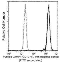
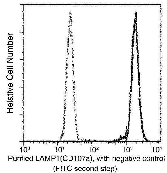
- Flow cytometric analysis of Human LAMP1 on Jurkat cells. Cells were treated according to manufacturer’s manual, stained with LAMP1 Recombinant Rabbit Monoclonal Antibody (107) (Product # MA5-29385), then a FITC-conjugated Secondary antibody. The histograms were derived from gated events with the forward and side light-scatter characteristics of intact cells.
Supportive validation
- Submitted by
- Invitrogen Antibodies (provider)
- Main image

- Experimental details
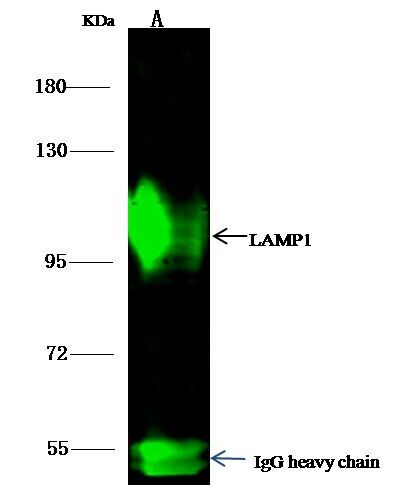
- LAMP1 Immunoprecipitation using: Lane A: 0.5 mg Jurkat Whole Cell Lysate 1 µL with LAMP1 Recombinant Rabbit Monoclonal Antibody (107) (Product # MA5-29385) and 15 µL of 50 % Protein G agarose. Primary antibody: LAMP1 Recombinant Rabbit Monoclonal Antibody (107), at 1:500 dilution. Secondary antibody: Dylight 800-labeled antibody to rabbit IgG (H+L), at 1:5,000 dilution. Developed using the Odyssey technique. Performed under reducing conditions. Predicted band size: 45 kDa. Observed band size: 113 kDa.
- Submitted by
- Invitrogen Antibodies (provider)
- Main image
- Experimental details
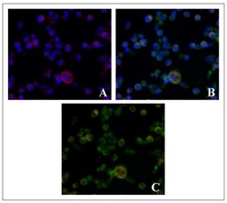
- Fluorescent microscopic images showing colocalization of lysosomal markers LAMP1 and BODIPY 1 in Ehrlich ascitic carcinoma cells: ( A )--BODIPY 1 (red) and nuclei (blue), ( B )--LAMP1 (green) and nuclei (blue), ( C )--overlapping BODIPY 1 and LAMP1 fluorescent staining. Magnification 40x.
