Explore
 Validate
 Learn
Learn Western blot
Antibody data
- Antibody Data
- Antigen structure
- References [0]
- Comments [0]
- Validations
- Western blot [2]
- Immunocytochemistry [2]
- Immunohistochemistry [6]
Submit
Validation data
Reference
Comment
Report error
- Product number
- GTX106424 - Provider product page

- Provider
- GeneTex
- Proper citation
- GeneTex Cat#GTX106424, RRID:AB_1240645
- Product name
- Coronin 1A antibody [C3], C-term
- Antibody type
- Polyclonal
- Reactivity
- Human, Mouse, Rat
- Host
- Rabbit
No comments: Submit comment
Supportive validation
- Submitted by
- GeneTex (provider)
- Main image
- Experimental details
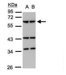
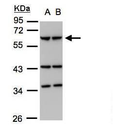
- Sample (30?g whole cell lysate)A:293TB:A431(GTX27909) 7.5% SDS PAGEGTX106424 diluted at 1:1000
- Validation comment
- WB
- Submitted by
- GeneTex (provider)
- Main image
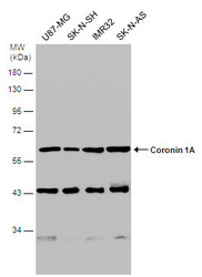
- Experimental details
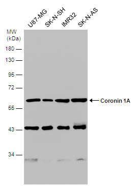
- Various whole cell extracts (30 ?g) were separated by 10% SDS-PAGE, and the membrane was blotted with Coronin 1A antibody [C3], C-term (GTX106424) diluted at 1:1000.
Supportive validation
- Submitted by
- GeneTex (provider)
- Main image
- Experimental details
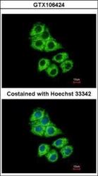
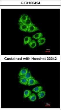
- Immunofluorescence analysis of methanol-fixed Hep G2, using Coronin 1A(GTX106424) antibody at 1:500 dilution.
- Submitted by
- GeneTex (provider)
- Main image

- Experimental details
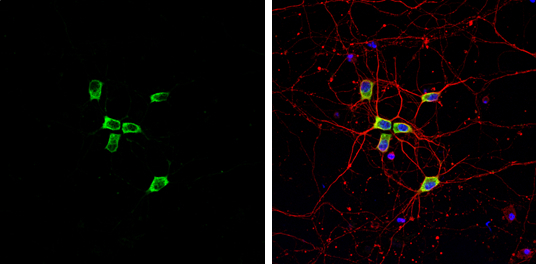
- Coronin 1A antibody [C3], C-term detects Coronin 1A protein by immunofluorescent analysis.Sample: DIV9 rat E18 primary cortical neuron cells were fixed in 4% paraformaldehyde at RT for 15 min.Green: Coronin 1A stained by Coronin 1A antibody [C3], C-term (GTX106424) diluted at 1:500.Red: beta Tubulin 3/ Tuj1, stained by beta Tubulin 3/ Tuj1 antibody [GT1338] (GTX631831) diluted at 1:500.Blue: Fluoroshield with DAPI (GTX30920).
Supportive validation
- Submitted by
- GeneTex (provider)
- Main image
- Experimental details
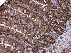
- Immunohistochemical analysis of paraffin-embedded human gastric cancer, using Coronin 1A(GTX106424) antibody at 1:100 dilution.
- Submitted by
- GeneTex (provider)
- Main image
- Experimental details
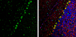
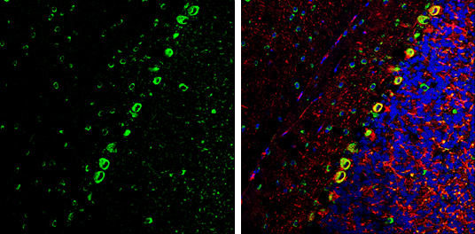
- Coronin 1A antibody [C3], C-term detects Coronin 1A Protein expression by immunohistochemical analysis.Sample: Frozen-sectioned adult mouse cerebellum. Green: Coronin 1A stained by Coronin 1A antibody [C3], C-term (GTX106424) diluted at 1:250.Red: NF-H, stained by NF-H antibody [GT114] (GTX634289) diluted at 1:500.Blue: Fluoroshield with DAPI (GTX30920).
- Submitted by
- GeneTex (provider)
- Main image
- Experimental details
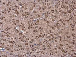
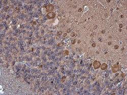
- Coronin 1A antibody [C3], C-term detects Coronin 1A protein at cytoplasm in mouse brain by immunohistochemical analysis. Sample: Paraffin-embedded mouse brain. Coronin 1A antibody [C3], C-term (GTX106424) diluted at 1:500.
- Submitted by
- GeneTex (provider)
- Main image
- Experimental details
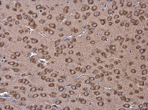
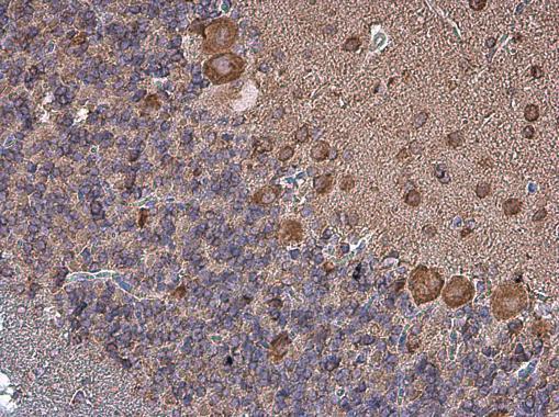
- Coronin 1A antibody [C3], C-term detects Coronin 1A protein at cytoplasm in rat brain by immunohistochemical analysis. Sample: Paraffin-embedded rat brain. Coronin 1A antibody [C3], C-term (GTX106424) diluted at 1:500.
- Submitted by
- GeneTex (provider)
- Main image
- Experimental details
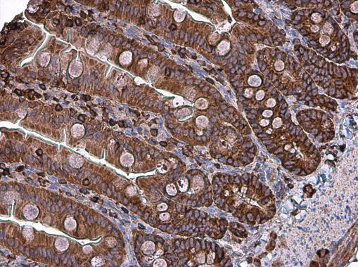
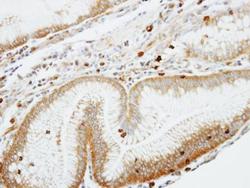
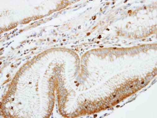
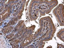
- Coronin 1A antibody [C3], C-term detects Coronin 1A protein at cytoplasm in mouse duodenum by immunohistochemical analysis. Sample: Paraffin-embedded mouse duodenum. Coronin 1A antibody [C3], C-term (GTX106424) diluted at 1:500.
- Submitted by
- GeneTex (provider)
- Main image
- Experimental details
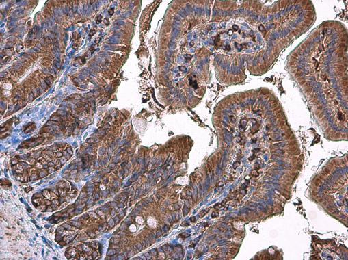
- Coronin 1A antibody [C3], C-term detects Coronin 1A protein at cytoplasm in rat intestine by immunohistochemical analysis. Sample: Paraffin-embedded rat intestine. Coronin 1A antibody [C3], C-term (GTX106424) diluted at 1:500.