Explore
 Validate
 Learn
Learn Western blot
 Immunocytochemistry
 Immunoprecipitation
Antibody data
- Antibody Data
- Antigen structure
- References [4]
- Comments [0]
- Validations
- Western blot [3]
- Immunocytochemistry [2]
- Immunohistochemistry [1]
Submit
Validation data
Reference
Comment
Report error
- Product number
- GTX105624 - Provider product page

- Provider
- GeneTex
- Proper citation
- GeneTex Cat#GTX105624, RRID:AB_1950645
- Product name
- Cytokeratin 18 antibody [N2C2], Internal
- Antibody type
- Polyclonal
- Reactivity
- Human, Mouse
- Host
- Rabbit
Submitted references Proteomic analysis of evodiamine-induced cytotoxicity in thyroid cancer cells.
High-efficiency cellular reprogramming with microfluidics.
Resources for the Comprehensive Discovery of Functional RNA Elements.
Klf5 deletion promotes Pten deletion-initiated luminal-type mouse prostate tumors through multiple oncogenic signaling pathways.
Yu HI, Chou HC, Su YC, Lin LH, Lu CH, Chuang HH, Tsai YT, Liao EC, Wei YS, Yang YT, Lee YR, Chan HL
Journal of pharmaceutical and biomedical analysis 2018 Oct 25;160:344-350
Journal of pharmaceutical and biomedical analysis 2018 Oct 25;160:344-350
High-efficiency cellular reprogramming with microfluidics.
Luni C, Giulitti S, Serena E, Ferrari L, Zambon A, Gagliano O, Giobbe GG, Michielin F, Knöbel S, Bosio A, Elvassore N
Nature methods 2016 May;13(5):446-52
Nature methods 2016 May;13(5):446-52
Resources for the Comprehensive Discovery of Functional RNA Elements.
Sundararaman B, Zhan L, Blue SM, Stanton R, Elkins K, Olson S, Wei X, Van Nostrand EL, Pratt GA, Huelga SC, Smalec BM, Wang X, Hong EL, Davidson JM, Lécuyer E, Graveley BR, Yeo GW
Molecular cell 2016 Mar 17;61(6):903-13
Molecular cell 2016 Mar 17;61(6):903-13
Klf5 deletion promotes Pten deletion-initiated luminal-type mouse prostate tumors through multiple oncogenic signaling pathways.
Xing C, Ci X, Sun X, Fu X, Zhang Z, Dong EN, Hao ZZ, Dong JT
Neoplasia (New York, N.Y.) 2014 Nov;16(11):883-99
Neoplasia (New York, N.Y.) 2014 Nov;16(11):883-99
No comments: Submit comment
Enhanced validation
Supportive validation
- Submitted by
- GeneTex (provider)
- Enhanced method
- Genetic validation
- Main image
- Experimental details
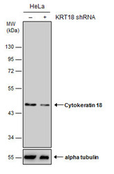
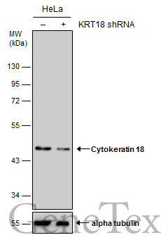
- Non-transfected (¡V) and transfected (+) HeLa whole cell extracts (15 ?g) were separated by 10% SDS-PAGE, and the membrane was blotted with Cytokeratin 18 antibody [N2C2], Internal (GTX105624) diluted at 1:2000.
Supportive validation
- Submitted by
- GeneTex (provider)
- Main image
- Experimental details
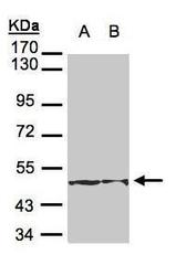
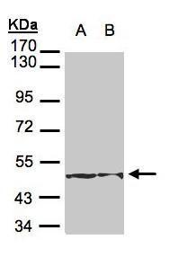
- Sample(30 £gg of whole cell lysate)A:H1299B:Hep G2(GTX27900)7.5% SDS PAGEGTX105624 diluted at 1:3000
- Submitted by
- GeneTex (provider)
- Main image

- Experimental details
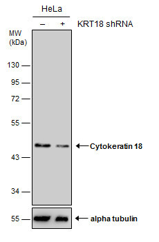
- Non-transfected (¡V) and transfected (+) HeLa whole cell extracts (15 ?g) were separated by 10% SDS-PAGE, and the membrane was blotted with Cytokeratin 18 antibody [N2C2], Internal (GTX105624) diluted at 1:2000.
Supportive validation
- Submitted by
- GeneTex (provider)
- Main image
- Experimental details
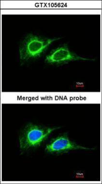
- Immunofluorescence analysis of paraformaldehyde-fixed HeLa, using Cytokeratin 18 (GTX105624) antibody at 1:200 dilution.
- Submitted by
- GeneTex (provider)
- Main image

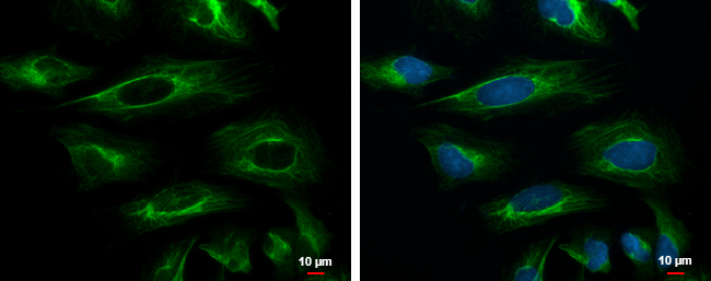
- Experimental details
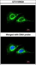
- Cytokeratin 18 antibody [N2C2], Internal detects Cytokeratin 18 protein at cytoskeleton by immunofluorescent analysis.Sample: HeLa cells were fixed in ice-cold MeOH for 5 min.Green: Cytokeratin 18 protein stained by Cytokeratin 18 antibody [N2C2], Internal (GTX105624) diluted at 1:200.Blue: Hoechst 33342 staining.Scale bar = 10 £gm.
Supportive validation
- Submitted by
- GeneTex (provider)
- Main image

- Experimental details
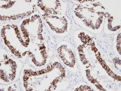
- Immunohistochemical analysis of paraffin-embedded human endo mitral ovarian cancer, using KRT18(GTX105624) antibody at 1:100 dilution.