 Explore
 Validate
 Learn
Learn Western blot
Antibody data
- Antibody Data
- Antigen structure
- References [2]
- Comments [0]
- Validations
- Western blot [2]
- Immunocytochemistry [1]
- Immunohistochemistry [1]
Submit
Validation data
Reference
Comment
Report error
- Product number
- AF7619 - Provider product page

- Provider
- R&D Systems
- Product name
- Human Cytokeratin 18 Antibody
- Antibody type
- Polyclonal
- Description
- Antigen Affinity-purified. Detects human Cytokeratin 18 in direct ELISAs and Western blots. In direct ELISAs, less than 1% cross-reactivity with recombinant human Cytokeratin 14 (KRT14) is observed.
- Reactivity
- Human
- Host
- Sheep
- Conjugate
- Unconjugated
- Antigen sequence
P05783- Isotype
- IgG
- Vial size
- 100 ug
- Concentration
- LYOPH
- Storage
- Use a manual defrost freezer and avoid repeated freeze-thaw cycles. 12 months from date of receipt, -20 to -70 °C as supplied. 1 month, 2 to 8 °C under sterile conditions after reconstitution. 6 months, -20 to -70 °C under sterile conditions after reconstitution.
Submitted references TFAP2C- and p63-Dependent Networks Sequentially Rearrange Chromatin Landscapes to Drive Human Epidermal Lineage Commitment.
Retinoic acid and BMP4 cooperate with p63 to alter chromatin dynamics during surface epithelial commitment.
Li L, Wang Y, Torkelson JL, Shankar G, Pattison JM, Zhen HH, Fang F, Duren Z, Xin J, Gaddam S, Melo SP, Piekos SN, Li J, Liaw EJ, Chen L, Li R, Wernig M, Wong WH, Chang HY, Oro AE
Cell stem cell 2019 Feb 7;24(2):271-284.e8
Cell stem cell 2019 Feb 7;24(2):271-284.e8
Retinoic acid and BMP4 cooperate with p63 to alter chromatin dynamics during surface epithelial commitment.
Pattison JM, Melo SP, Piekos SN, Torkelson JL, Bashkirova E, Mumbach MR, Rajasingh C, Zhen HH, Li L, Liaw E, Alber D, Rubin AJ, Shankar G, Bao X, Chang HY, Khavari PA, Oro AE
Nature genetics 2018 Dec;50(12):1658-1665
Nature genetics 2018 Dec;50(12):1658-1665
No comments: Submit comment
Supportive validation
- Submitted by
- R&D Systems (provider)
- Main image

- Experimental details
- Detection of Human Cytokeratin 18 by Western Blot. Western blot shows lysates of HeLa human cervical epithelial carcinoma cell line and A431 human epithelial carcinoma cell line. PVDF membrane was probed with 0.1 µg/mL of Sheep Anti-Human Cytokeratin 18 Antigen Affinity-purified Polyclonal Antibody (Catalog # AF7619) followed by HRP-conjugated Anti-Sheep IgG Secondary Antibody (Catalog # HAF016). A specific band was detected for Cytokeratin 18 at approximately 46 kDa (as indicated). This experiment was conducted under reducing conditions and using Immunoblot Buffer Group 1.

- Submitted by
- R&D Systems (provider)
- Main image

- Experimental details
- Detection of Human Cytokeratin 18 by Simple WesternTM. Simple Western lane view shows lysates of HeLa human cervical epithelial carcinoma cell line and A431 human epithelial carcinoma cell line, loaded at 0.2 mg/mL. A specific band was detected for Cytokeratin 18 at approximately 57 kDa (as indicated) using 1 µg/mL of Sheep Anti-Human Cytokeratin 18 Antigen Affinity-purified Polyclonal Antibody (Catalog # AF7619) followed by 1:50 dilution of HRP-conjugated Anti-Sheep IgG Secondary Antibody (Catalog # HAF016). This experiment was conducted under reducing conditions and using the 12-230 kDa separation system.

Supportive validation
- Submitted by
- R&D Systems (provider)
- Main image

- Experimental details
- Cytokeratin 18 in HeLa Human Cell Line. Cytokeratin 18 was detected in immersion fixed HeLa human cervical epithelial carcinoma cell line using Sheep Anti-Human Cytokeratin 18 Antigen Affinity-purified Polyclonal Antibody (Catalog # AF7619) at 5 µg/mL for 3 hours at room temperature. Cells were stained using the NorthernLights™ 557-conjugated Anti-Sheep IgG Secondary Antibody (red; Catalog # NL010) and counterstained with DAPI (blue). Specific staining was localized to cytoskeletal fibers. View our protocol for Fluorescent ICC Staining of Cells on Coverslips.

Supportive validation
- Submitted by
- R&D Systems (provider)
- Main image
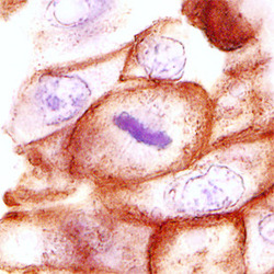
- Experimental details
- Cytokeratin 18 in Human Breast Cancer Tissue. Cytokeratin 18 was detected in immersion fixed paraffin-embedded sections of human breast cancer tissue using Sheep Anti-Human Cytokeratin 18 Antigen Affinity-purified Polyclonal Antibody (Catalog # AF7619) at 3 µg/mL overnight at 4 °C. Before incubation with the primary antibody, tissue was subjected to heat-induced epitope retrieval using Antigen Retrieval Reagent-Basic (Catalog # CTS013). Tissue was stained using the Anti-Sheep HRP-DAB Cell & Tissue Staining Kit (brown; Catalog # CTS019) and counterstained with hematoxylin (blue). Specific staining was localized to intermediate filaments. View our protocol for Chromogenic IHC Staining of Paraffin-embedded Tissue Sections.
