 Explore
 Validate
 Learn
Learn Western blot
 ELISA
ELISAAntibody data
- Antibody Data
- Antigen structure
- References [2]
- Comments [0]
- Validations
- Western blot [1]
- Immunohistochemistry [1]
- Flow cytometry [1]
Submit
Validation data
Reference
Comment
Report error
- Product number
- ABIN453171 - Provider product page

- Provider
- antibodies-online
- Product name
- anti-Lymphotoxin alpha (TNF Superfamily, Member 1) (LTA) (Center) antibody
- Antibody type
- Polyclonal
- Antigen
- KLH conjugated synthetic peptide selected from the Center region of human LTA
- Reactivity
- Human
- Host
- Rabbit
- Vial size
- 0.1 mg
- Concentration
- 0.25 mg/ml
- Storage
- Store the antibody undiluted at 2-8°C for one month or (in aliquots) at -20°C for longer. Avoid repeated freezing and thawing. Shelf life: one year from despatch.
Submitted references N-linked sugar chain structure of recombinant human lymphotoxin produced by CHO cells: the functional role of carbohydrate as to its lectin-like character and clearance velocity.
Effects of the human immunodeficiency virus type 1 Tat protein on the expression of inflammatory cytokines.
Fukushima K, Watanabe H, Takeo K, Nomura M, Asahi T, Yamashita K
Archives of biochemistry and biophysics 1993 Jul;304(1):144-53
Archives of biochemistry and biophysics 1993 Jul;304(1):144-53
Effects of the human immunodeficiency virus type 1 Tat protein on the expression of inflammatory cytokines.
Buonaguro L, Barillari G, Chang HK, Bohan CA, Kao V, Morgan R, Gallo RC, Ensoli B
Journal of virology 1992 Dec;66(12):7159-67
Journal of virology 1992 Dec;66(12):7159-67
No comments: Submit comment
Supportive validation
- Submitted by
- antibodies-online (provider)
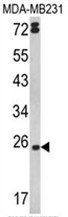
- Main image

- Experimental details
- Western blot analysis of LTA Antibody (Center) (AP17540PU-N) in MDA-MB231 cell line lysates (35ug/lane). LTA (arrow) was detected using the purified Pab.
Supportive validation
- Submitted by
- antibodies-online (provider)
- Main image

- Experimental details
- Formalin-fixed and paraffin-embedded human hepatocarcinoma reacted with LTA Antibody (Center), which was peroxidase-conjugated to the secondary antibody, followed by DAB staining.

Supportive validation
- Submitted by
- antibodies-online (provider)
- Main image

- Experimental details
- Flow Cytometric analysis of HepG2 cells using LTA Antibody (Center)(bottom histogram) compared to a negative control cell (top histogram). FITC-conjugated Goat-anti-Rabbit secondary antibodies were used for the analysis.
