Explore
 Validate
 Learn
Learn Western blot
 ELISA
ELISAAntibody data
- Antibody Data
- Antigen structure
- References [2]
- Comments [0]
- Validations
- Western blot [1]
- Immunocytochemistry [1]
- Immunohistochemistry [1]
Submit
Validation data
Reference
Comment
Report error
- Product number
- ABIN954045 - Provider product page

- Provider
- antibodies-online
- Product name
- anti-Protocadherin beta 3 (PCDHB3) (N-Term) antibody
- Antibody type
- Polyclonal
- Antigen
- Other
- Reactivity
- Human
- Host
- Rabbit
- Vial size
- 0.1 mg
Submitted references Protocadherins.
Comparative DNA sequence analysis of mouse and human protocadherin gene clusters.
Frank M, Kemler R
Current opinion in cell biology 2002 Oct;14(5):557-62
Current opinion in cell biology 2002 Oct;14(5):557-62
Comparative DNA sequence analysis of mouse and human protocadherin gene clusters.
Wu Q, Zhang T, Cheng JF, Kim Y, Grimwood J, Schmutz J, Dickson M, Noonan JP, Zhang MQ, Myers RM, Maniatis T
Genome research 2001 Mar;11(3):389-404
Genome research 2001 Mar;11(3):389-404
No comments: Submit comment
Supportive validation
- Submitted by
- antibodies-online (provider)
- Main image
- Experimental details
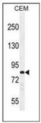
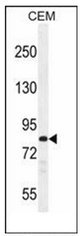
- Western blot analysis of PCDHB3 Antibody (N-term) Cat.-No AP53211PU-N in CEM cell line lysates (35ug/lane). This demonstrates the PCDHB3 antibody detected the PCDHB3 protein (arrow).
Supportive validation
- Submitted by
- antibodies-online (provider)
- Main image
- Experimental details
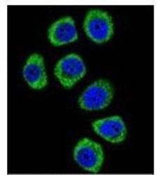
- Confocal immunofluorescent analysis of PCDHB3 Antibody (N-term) Cat.-No AP53211PU-N with U-251MG cell followed by Alexa Fluor 488-conjugated goat anti-rabbit lgG (green). DAPI was used to stain the cell nuclear (blue).

Supportive validation
- Submitted by
- antibodies-online (provider)
- Main image

- Experimental details
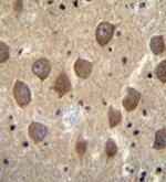
- Immunohistochemistry analysis in formalin fixed and paraffin embedded human brain tissue reacted with PCDHB3 Antibody (N-term) Cat.-No AP53211PU-N followed by peroxidase conjugation of the secondary antibody and DAB staining.