 Explore
 Validate
 Learn
Learn Western blot
Antibody data
- Antibody Data
- Antigen structure
- References [3]
- Comments [0]
- Validations
- Western blot [1]
- Immunocytochemistry [1]
- Other assay [1]
Submit
Validation data
Reference
Comment
Report error
- Product number
- MA5-23720 - Provider product page

- Provider
- Invitrogen Antibodies
- Product name
- TNF alpha Monoclonal Antibody (28401)
- Antibody type
- Monoclonal
- Antigen
- Recombinant full-length protein
- Description
- In sandwich ELISAs, less than 0.05% cross-reactivity with recombinant human TNF- beta, recombinant mouse TNF- alpha, recombinant rat TNF- alpha, and recombinant porcine TNF- alpha is observed.
- Antibody clone number
- 28401
- Concentration
- 0.5 mg/mL
Submitted references The Ameliorative Role of Eugenol against Silver Nanoparticles-Induced Hepatotoxicity in Male Wistar Rats.
Intact Transition Epitope Mapping - Targeted High-Energy Rupture of Extracted Epitopes (ITEM-THREE).
Neutrophil Microvesicles from Healthy Control and Rheumatoid Arthritis Patients Prevent the Inflammatory Activation of Macrophages.
Yousef HN, Ibraheim SS, Ramadan RA, Aboelwafa HR
Oxidative medicine and cellular longevity 2022;2022:3820848
Oxidative medicine and cellular longevity 2022;2022:3820848
Intact Transition Epitope Mapping - Targeted High-Energy Rupture of Extracted Epitopes (ITEM-THREE).
Danquah BD, Röwer C, Opuni KM, El-Kased R, Frommholz D, Illges H, Koy C, Glocker MO
Molecular & cellular proteomics : MCP 2019 Aug;18(8):1543-1555
Molecular & cellular proteomics : MCP 2019 Aug;18(8):1543-1555
Neutrophil Microvesicles from Healthy Control and Rheumatoid Arthritis Patients Prevent the Inflammatory Activation of Macrophages.
Rhys HI, Dell'Accio F, Pitzalis C, Moore A, Norling LV, Perretti M
EBioMedicine 2018 Mar;29:60-69
EBioMedicine 2018 Mar;29:60-69
No comments: Submit comment
Supportive validation
- Submitted by
- Invitrogen Antibodies (provider)
- Main image
- Experimental details
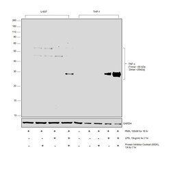
- Western Blot was performed using Anti-TNF alpha Monoclonal Antibody (28401) (Product # MA5-23720) and a 25 kDa band corresponding to TNF alpha was detected in lipopolysaccharide treated macrophages (differentiated from both THP-1 and U-937). Whole cell extracts (30 µg lysate) of untreated macrophages differentiated from U-937 (Lane 1), treated with PTI (Lane 2), stimulated with LPS (Lane 3), stimulated with LPS and subsequently treated with PTI (Lane 4), and THP-1 control (Lane 5), untreated, THP-1 differentiated macrophage (Lane 6), differentiated macrophage treated with PTI (Lane 7), THP-1 macrophage stimulated with LPS (Lane 8), THP-1 macrophage stimulated with LPS and then treated with PTI (Lane 9) were electrophoresed using NuPAGE™ 12% Bis-Tris Protein Gel (Product # NP0341BOX). Resolved proteins were then transferred onto a Nitrocellulose membrane (Product # IB23001) by iBlot® 2 Dry Blotting System (Product # IB21001). The Blot was probed with the primary antibody (1:1000 dilution) and detected by chemiluminescence with Goat anti-Mouse IgG (H+L) Superclonal™ Recombinant Secondary Antibody, HRP (Product # A28177, 1:10000 dilution) using the iBright FL 1000 (Product # A32752). Chemiluminescent detection was performed using SuperSignal™ West Dura Extended Duration Substrate (Product # 34076).

Supportive validation
- Submitted by
- Invitrogen Antibodies (provider)
- Main image
- Experimental details
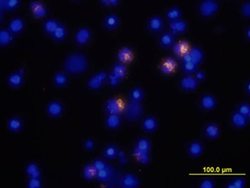
- Immunocytochemical analysis of TNF-a was detected in immersion fixed human peripheral blood mononuclear cells (PBMCs) stimulated with LPS and monensin using mouse Anti-human TNF-a Monoclonal Antibody (Product # MA5-23720) at 10 µg/mL for 3 hours at room temperature. Cells were stained using the 557-conjugated Anti-mouse IgG Secondary Antibody (yello and counterstained with DAPI (blue).

Supportive validation
- Submitted by
- Invitrogen Antibodies (provider)
- Main image
- Experimental details
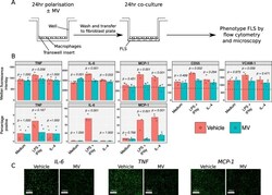
- Fig. 5 Effect of neutrophil MVs on macrophage-FLS co-culture. A) Experiment scheme. Monocyte-derived macrophages were treated with 10 ng/mL LPS and 20 ng/mL IFN-gamma, 50 ng/mL IL-4 or vehicle for 24 h, in the presence of 3 x 10 6 MV/mL or vehicle. Macrophages were washed and co-cultured with FLS for a further 24 h, after which time FLS were immunophenotyped by flow cytometry. The experiment was repeated with 3 different macrophage donors and the mean across the donors was taken for each replicate. B) Antigen expression data for each FLS donor (mean expression across co-culture with 3 different macrophage donors) where dotted lines indicate median fluorescence of isotype-matched control samples. C) Confirmatory immunofluorescence images of IL-6, TNF-alpha and MCP-1 expression after co-culture with macrophages after classical activation, in the presence of 3 x 10 6 MV/mL or vehicle. Data in B analysed with separate two-way ANOVA with Holm-Sidak post-hoc tests for each antigen. Fig. 5
