Explore
 Validate
 Learn
Learn Western blot
 ELISA
ELISA Immunohistochemistry
Antibody data
- Antibody Data
- Antigen structure
- References [0]
- Comments [0]
- Validations
- Western blot [1]
- Immunocytochemistry [2]
- Immunohistochemistry [14]
Submit
Validation data
Reference
Comment
Report error
- Product number
- LS-C782289 - Provider product page

- Provider
- LSBio
- Product name
- BAG6 / G3 / Scythe Antibody LS-C782289
- Antibody type
- Polyclonal
- Description
- Antigen Affinity purification
- Reactivity
- Human, Mouse, Rat
- Host
- Rabbit
- Isotype
- IgG
- Storage
- After reconstitution, store at 4°C for up to 1 month. Long-term: aliquot and store at -20°C. Avoid freeze-thaws cycles.
No comments: Submit comment
Supportive validation
- Submitted by
- LSBio (provider)
- Enhanced method
- Genetic validation
- Main image
- Experimental details
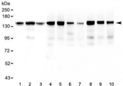
- Western blot testing of 1) human HeLa, 2) human ThP-1, 3) human U-87 MG, 4) rat brain, 5) rat heart, 6) rat lung, 7) rat liver, 8) mouse brain, 9) mouse heart and 10) mouse lung lysate with BAG6 antibody at 0.5ug/ml. Predicted molecular weight ~119 kDa but observed at 150-170 kDa.

Supportive validation
- Submitted by
- LSBio (provider)
- Enhanced method
- Genetic validation
- Main image
- Experimental details
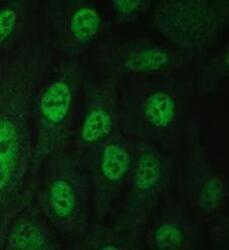
- IF/ICC staining of FFPE human U-2 OS cells with BAG6 antibody (green) at 2ug/ml. HIER: steam section in pH6 citrate buffer for 20 min.

- Submitted by
- LSBio (provider)
- Main image

- Experimental details
- IF/ICC staining of FFPE human U-2 OS cells with BAG6 antibody (green) at 2ug/ml. HIER: steam section in pH6 citrate buffer for 20 min.

Enhanced validation
- Submitted by
- LSBio (provider)
- Enhanced method
- Genetic validation
- Main image

- Experimental details
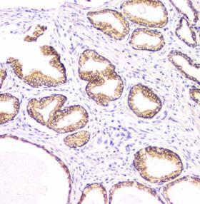
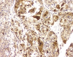
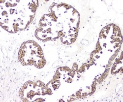
- IHC staining of FFPE human prostate cancer with BAG6 antibody at 1ug/ml. HIER: boil tissue sections in pH6, 10mM citrate buffer, for 10-20 min and allow to cool before testing.
- Submitted by
- LSBio (provider)
- Enhanced method
- Genetic validation
- Main image

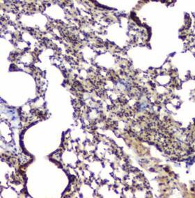
- Experimental details
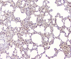
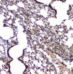
- IHC staining of FFPE mouse lung with BAG6 antibody at 1ug/ml. HIER: boil tissue sections in pH6, 10mM citrate buffer, for 10-20 min and allow to cool before testing.

- Submitted by
- LSBio (provider)
- Enhanced method
- Genetic validation
- Main image

- Experimental details
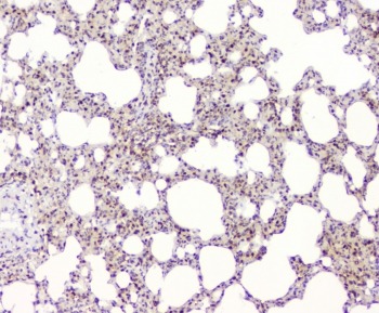
- IHC staining of FFPE rat lung with BAG6 antibody at 1ug/ml. HIER: boil tissue sections in pH6, 10mM citrate buffer, for 10-20 min and allow to cool before testing.

- Submitted by
- LSBio (provider)
- Enhanced method
- Genetic validation
- Main image

- Experimental details
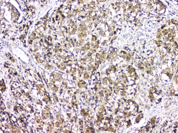
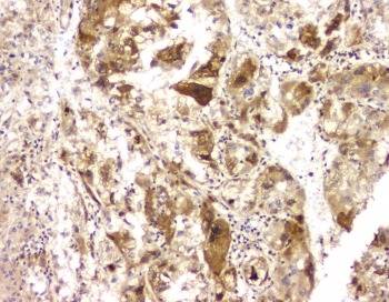
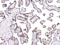
- IHC staining of FFPE human liver cancer with BAG6 antibody at 1ug/ml. HIER: boil tissue sections in pH6, 10mM citrate buffer, for 10-20 min and allow to cool before testing.

- Submitted by
- LSBio (provider)
- Enhanced method
- Genetic validation
- Main image

- Experimental details
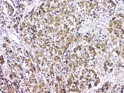
- IHC staining of FFPE human stomach cancer with BAG6 antibody at 1ug/ml. HIER: boil tissue sections in pH6, 10mM citrate buffer, for 10-20 min and allow to cool before testing.
- Submitted by
- LSBio (provider)
- Enhanced method
- Genetic validation
- Main image

- Experimental details
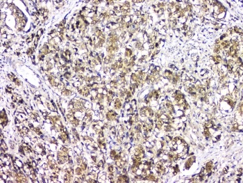
- IHC staining of FFPE human ovarian cancer with BAG6 antibody at 1ug/ml. HIER: boil tissue sections in pH6, 10mM citrate buffer, for 10-20 min and allow to cool before testing.

- Submitted by
- LSBio (provider)
- Enhanced method
- Genetic validation
- Main image

- Experimental details
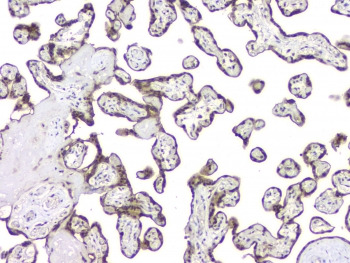
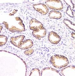
- IHC staining of FFPE human placenta with BAG6 antibody at 1ug/ml. HIER: boil tissue sections in pH6, 10mM citrate buffer, for 10-20 min and allow to cool before testing.
- Submitted by
- LSBio (provider)
- Main image
- Experimental details
- IHC staining of FFPE rat lung with BAG6 antibody at 1ug/ml. HIER: boil tissue sections in pH6, 10mM citrate buffer, for 10-20 min and allow to cool before testing.
- Submitted by
- LSBio (provider)
- Main image
- Experimental details
- IHC staining of FFPE human liver cancer with BAG6 antibody at 1ug/ml. HIER: boil tissue sections in pH6, 10mM citrate buffer, for 10-20 min and allow to cool before testing.
- Submitted by
- LSBio (provider)
- Main image
- Experimental details
- IHC staining of FFPE human stomach cancer with BAG6 antibody at 1ug/ml. HIER: boil tissue sections in pH6, 10mM citrate buffer, for 10-20 min and allow to cool before testing.
- Submitted by
- LSBio (provider)
- Main image
- Experimental details
- IHC staining of FFPE human ovarian cancer with BAG6 antibody at 1ug/ml. HIER: boil tissue sections in pH6, 10mM citrate buffer, for 10-20 min and allow to cool before testing.

- Submitted by
- LSBio (provider)
- Main image
- Experimental details
- IHC staining of FFPE human placenta with BAG6 antibody at 1ug/ml. HIER: boil tissue sections in pH6, 10mM citrate buffer, for 10-20 min and allow to cool before testing.

- Submitted by
- LSBio (provider)
- Main image
- Experimental details
- IHC staining of FFPE human prostate cancer with BAG6 antibody at 1ug/ml. HIER: boil tissue sections in pH6, 10mM citrate buffer, for 10-20 min and allow to cool before testing.

- Submitted by
- LSBio (provider)
- Main image
- Experimental details
- IHC staining of FFPE mouse lung with BAG6 antibody at 1ug/ml. HIER: boil tissue sections in pH6, 10mM citrate buffer, for 10-20 min and allow to cool before testing.