AF1022
antibody from Novus Biologicals
Targeting: PDCD1LG2
B7-DC, bA574F11.2, Btdc, CD273, PD-L2, PDL2
Antibody data
- Antibody Data
- Antigen structure
- References [3]
- Comments [0]
- Validations
- Immunohistochemistry [1]
Submit
Validation data
Reference
Comment
Report error
- Product number
- AF1022 - Provider product page

- Provider
- Novus Biologicals
- Product name
- Goat Polyclonal PD-L2/B7-DC/PDCD1LG2 Antibody
- Antibody type
- Polyclonal
- Description
- Immunogen affinity purified. Detects mouse PD-L2 in direct ELISAs and Western blots. In direct ELISAs and Western blots, approximately 20% cross-reactivity with recombinant human PD-L2 is observed.
- Reactivity
- Mouse
- Host
- Goat
- Isotype
- IgG
- Vial size
- 100 ug
- Concentration
- LYOPH
- Storage
- Use a manual defrost freezer and avoid repeated freeze-thaw cycles. 12 months from date of receipt, -20 to -70 degreesC as supplied. 1 month, 2 to 8 degreesC under sterile conditions after reconstitution. 6 months, -20 to -70 degreesC under sterile conditions after reconstitution.
Submitted references The Circadian Clock Controls Immune Checkpoint Pathway in Sepsis.
Anti-programmed cell death 1 antibody reduces CD4+PD-1+ T cells and relieves the lupus-like nephritis of NZB/W F1 mice.
Establishment of NOD-Pdcd1-/- mice as an efficient animal model of type I diabetes.
Deng W, Zhu S, Zeng L, Liu J, Kang R, Yang M, Cao L, Wang H, Billiar TR, Jiang J, Xie M, Tang D
Cell reports 2018 Jul 10;24(2):366-378
Cell reports 2018 Jul 10;24(2):366-378
Anti-programmed cell death 1 antibody reduces CD4+PD-1+ T cells and relieves the lupus-like nephritis of NZB/W F1 mice.
Kasagi S, Kawano S, Okazaki T, Honjo T, Morinobu A, Hatachi S, Shimatani K, Tanaka Y, Minato N, Kumagai S
Journal of immunology (Baltimore, Md. : 1950) 2010 Mar 1;184(5):2337-47
Journal of immunology (Baltimore, Md. : 1950) 2010 Mar 1;184(5):2337-47
Establishment of NOD-Pdcd1-/- mice as an efficient animal model of type I diabetes.
Wang J, Yoshida T, Nakaki F, Hiai H, Okazaki T, Honjo T
Proceedings of the National Academy of Sciences of the United States of America 2005 Aug 16;102(33):11823-8
Proceedings of the National Academy of Sciences of the United States of America 2005 Aug 16;102(33):11823-8
No comments: Submit comment
Supportive validation
- Submitted by
- Novus Biologicals (provider)
- Main image

- Experimental details
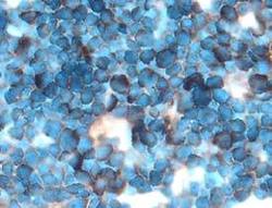
- PD-L2 in Mouse Thymus. PD-L2 was detected in perfusion fixed frozen sections of mouse thymus using 15 µg/mL Goat Anti-Mouse PD-L2 Antigen Affinity-purified Polyclonal Antibody (Catalog # AF1022) overnight at 4 °C. Tissue was stained with the Anti-Goat HRP-DAB Cell & Tissue Staining Kit (brown; Catalog # CTS008) and counterstained with hematoxylin (blue). View our protocol for Chromogenic IHC Staining of Frozen Tissue Sections.
 Explore
 Validate
 Learn
Learn Western blot
 Immunohistochemistry