 Explore
 Validate
 Learn
Learn Western blot
Antibody data
- Antibody Data
- Antigen structure
- References [2]
- Comments [0]
- Validations
- Western blot [3]
- Immunohistochemistry [1]
Submit
Validation data
Reference
Comment
Report error
- Product number
- AF4174 - Provider product page

- Provider
- R&D Systems
- Product name
- Human/Mouse/Rat FKBP12.6 Antibody
- Antibody type
- Polyclonal
- Description
- Antigen Affinity-purified. Detects human, mouse, and rat FKBP12.6 in Western blots. In Western blots, approximately 15% cross-reactivity with recombinant human FKBP12 and less than 1% cross-reactivity with recombinant human FKBP13 and FKBP25 is observed.
- Reactivity
- Human, Mouse, Rat
- Host
- Goat
- Conjugate
- Unconjugated
- Antigen sequence
P68106- Isotype
- IgG
- Vial size
- 100 ug
- Concentration
- LYOPH
- Storage
- Use a manual defrost freezer and avoid repeated freeze-thaw cycles. 12 months from date of receipt, -20 to -70 °C as supplied. 1 month, 2 to 8 °C under sterile conditions after reconstitution. 6 months, -20 to -70 °C under sterile conditions after reconstitution.
Submitted references Specific and Novel microRNAs Are Regulated as Response to Fungal Infection in Human Dendritic Cells.
Impact of hypoxia, simulated ischemia and reperfusion in HL-1 cells on the expression of FKBP12/FKBP12.6 and intracellular calcium dynamics.
Dix A, Czakai K, Leonhardt I, Schäferhoff K, Bonin M, Guthke R, Einsele H, Kurzai O, Löffler J, Linde J
Frontiers in microbiology 2017;8:270
Frontiers in microbiology 2017;8:270
Impact of hypoxia, simulated ischemia and reperfusion in HL-1 cells on the expression of FKBP12/FKBP12.6 and intracellular calcium dynamics.
Åström-Olsson K, Li L, Olofsson CS, Borén J, Öhlin H, Grip L
Biochemical and biophysical research communications 2012 Jun 15;422(4):732-8
Biochemical and biophysical research communications 2012 Jun 15;422(4):732-8
No comments: Submit comment
Supportive validation
- Submitted by
- R&D Systems (provider)
- Main image

- Experimental details
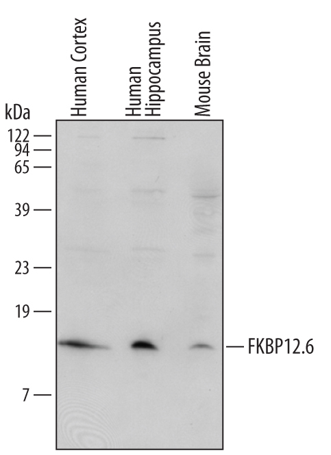
- Detection of Human/Mouse/Rat FKBP12.6 by Western Blot. Western blot shows lysates of human cortex, human hippocampus, and mouse brain tissues. PVDF membrane was probed with 1 µg/mL of Human/Mouse/Rat FKBP12.6 Antigen Affinity-purified Polyclonal Antibody (Catalog # AF4174) followed by HRP-conjugated Anti-Goat IgG Secondary Antibody (Catalog # HAF109). A specific band was detected for FKBP12.6 at approximately 13 kDa (as indicated). This experiment was conducted using Immunoblot Buffer Group 2.
- Submitted by
- R&D Systems (provider)
- Main image
- Experimental details
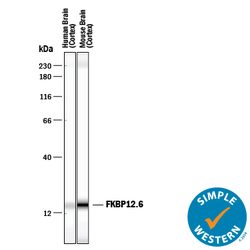
- Detection of Human and Mouse FKBP12.6 by Simple WesternTM. Simple Western lane view shows lysates of human brain (cortex) tissue and mouse brain (cortex) tissue, loaded at 0.2 mg/mL. A specific band was detected for FKBP12.6 at approximately 16 kDa (as indicated) using 50 µg/mL of Goat Anti-Human/Mouse/Rat FKBP12.6 Antigen Affinity-purified Polyclonal Antibody (Catalog # AF4174) followed by 1:50 dilution of HRP-conjugated Anti-Goat IgG Secondary Antibody (Catalog # HAF109). This experiment was conducted under reducing conditions and using the 12-230 kDa separation system.

- Submitted by
- R&D Systems (provider)
- Main image
- Experimental details
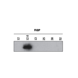
- Specificity of FKBP12.6 Shown by Western Blot. Western blot shows recombinant human FKBP 12, recombinant human FKBP 12.6, recombinant human FKBP 13, recombinant human FKBP 25, recombinant human FKBP 38, and recombinant human FKBP 52. PVDF membrane was probed with 1 µg/mL of Goat Anti-Human/Mouse/Rat FKBP12.6 Antigen Affinity-purified Polyclonal Antibody (Catalog # AF4174) followed by HRP-conjugated Anti-Goat IgG Secondary Antibody (Catalog # HAF017). A specific band was detected for FKBP12.6 at approximately 13 kDa (as indicated). This experiment was conducted under reducing conditions and using Immunoblot Buffer Group 1.

Supportive validation
- Submitted by
- R&D Systems (provider)
- Main image

- Experimental details
- FKBP12.6 in Human Brain. FKBP12.6 was detected in immersion fixed paraffin-embedded sections of human brain (caudate nucleus) using Goat Anti-Human/Mouse/Rat FKBP12.6 Antigen Affinity-purified Polyclonal Antibody (Catalog # AF4174) at 10 µg/mL for 1 hour at room temperature followed by incubation with the Anti-Goat IgG VisUCyte™ HRP Polymer Antibody (Catalog # VC004). Tissue was stained using DAB (brown) and counterstained with hematoxylin (blue). Specific staining was localized to cytoplasm in neurons. View our protocol for IHC Staining with VisUCyte HRP Polymer Detection Reagents.
