Explore
 Validate
 Learn
Learn Western blot
Antibody data
- Antibody Data
- Antigen structure
- References [5]
- Comments [0]
- Validations
- Western blot [2]
- Immunohistochemistry [3]
- Other assay [6]
Submit
Validation data
Reference
Comment
Report error
- Product number
- PA1-759 - Provider product page

- Provider
- Invitrogen Antibodies
- Product name
- Ubiquilin 1 Polyclonal Antibody
- Antibody type
- Polyclonal
- Antigen
- Synthetic peptide
- Description
- PA1-759 detects ubiquilin from hamster cells.
- Concentration
- 1 mg/mL
Submitted references GABA(A) Receptor-Stabilizing Protein Ubqln1 Affects Hyperexcitability and Epileptogenesis after Traumatic Brain Injury and in a Model of In Vitro Epilepsy in Mice.
Signature changes in ubiquilin expression in the R6/2 mouse model of Huntington's disease.
Effects of ubiquilin 1 on the unfolded protein response.
Ubiquilin 1 modulates amyloid precursor protein trafficking and Abeta secretion.
Identification of ubiquilin, a novel presenilin interactor that increases presenilin protein accumulation.
Kürten T, Ihbe N, Ueberbach T, Distler U, Sielaff M, Tenzer S, Mittmann T
International journal of molecular sciences 2022 Mar 31;23(7)
International journal of molecular sciences 2022 Mar 31;23(7)
Signature changes in ubiquilin expression in the R6/2 mouse model of Huntington's disease.
Safren N, Chang L, Dziki KM, Monteiro MJ
Brain research 2015 Feb 9;1597:37-46
Brain research 2015 Feb 9;1597:37-46
Effects of ubiquilin 1 on the unfolded protein response.
Lu A, Hiltunen M, Romano DM, Soininen H, Hyman BT, Bertram L, Tanzi RE
Journal of molecular neuroscience : MN 2009 May;38(1):19-30
Journal of molecular neuroscience : MN 2009 May;38(1):19-30
Ubiquilin 1 modulates amyloid precursor protein trafficking and Abeta secretion.
Hiltunen M, Lu A, Thomas AV, Romano DM, Kim M, Jones PB, Xie Z, Kounnas MZ, Wagner SL, Berezovska O, Hyman BT, Tesco G, Bertram L, Tanzi RE
The Journal of biological chemistry 2006 Oct 27;281(43):32240-53
The Journal of biological chemistry 2006 Oct 27;281(43):32240-53
Identification of ubiquilin, a novel presenilin interactor that increases presenilin protein accumulation.
Mah AL, Perry G, Smith MA, Monteiro MJ
The Journal of cell biology 2000 Nov 13;151(4):847-62
The Journal of cell biology 2000 Nov 13;151(4):847-62
No comments: Submit comment
Supportive validation
- Submitted by
- Invitrogen Antibodies (provider)
- Main image

- Experimental details
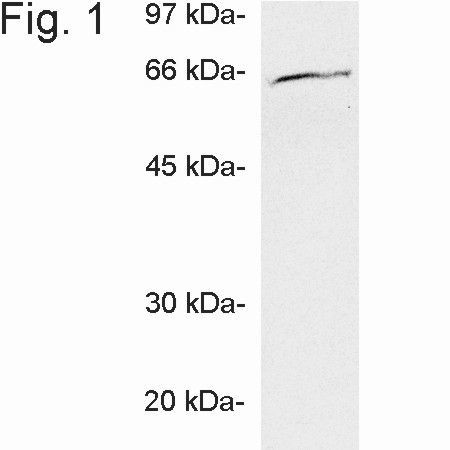
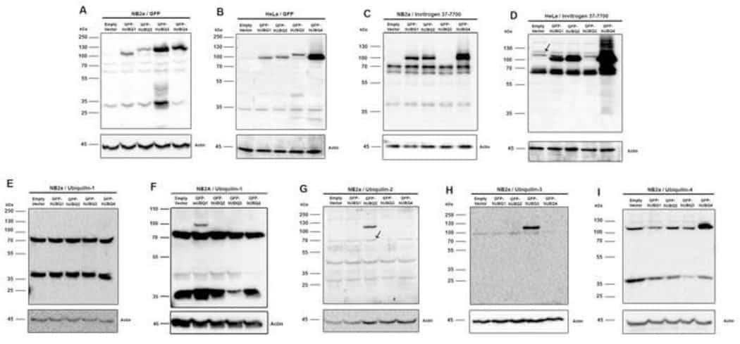
- Western blot of ubiquilin on CHO cell extract using Product # PA1-759.
- Submitted by
- Invitrogen Antibodies (provider)
- Main image

- Experimental details
- Western blot of ubiquilin on CHO cell extract using Product # PA1-759.

Supportive validation
- Submitted by
- Invitrogen Antibodies (provider)
- Main image
- Experimental details
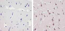
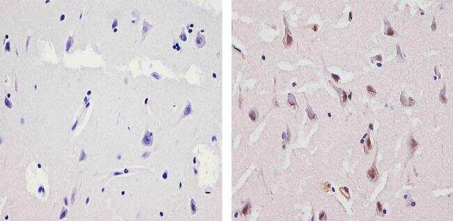
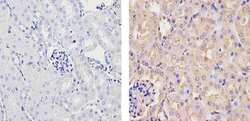
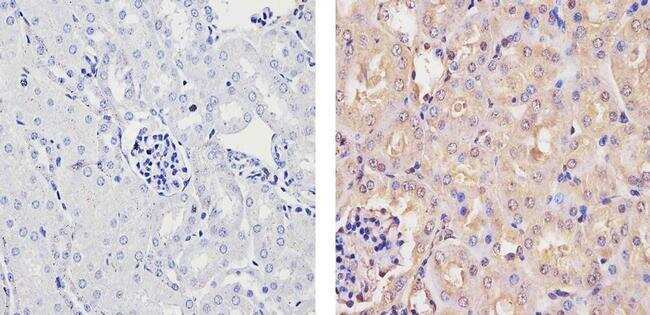
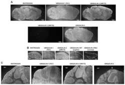
- Immunohistochemistry analysis of Ubiquilin showing staining in the nucleus and weak staining in the cytoplasm of paraffin-embedded human brain tissue (right) compared to a negative control without primary antibody (left). To expose target proteins, antigen retrieval was performed using 10mM sodium citrate (pH 6.0), microwaved for 8-15 min. Following antigen retrieval, tissues were blocked in 3% H2O2-methanol for 15 min at room temperature, washed with ddH2O and PBS, and then probed with a Ubiquilin Rabbit Polyclonal Antibody (Product # PA1-759) diluted in 3% BSA-PBS at a dilution of 1:100 for 1 hour at 37ºC in a humidified chamber. Tissues were washed extensively in PBST and detection was performed using an HRP-conjugated secondary antibody followed by colorimetric detection using a DAB kit. Tissues were counterstained with hematoxylin and dehydrated with ethanol and xylene to prep for mounting.
- Submitted by
- Invitrogen Antibodies (provider)
- Main image
- Experimental details
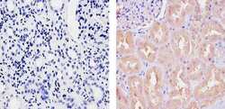
- Immunohistochemistry analysis of Ubiquilin showing staining in the cytoplasm and nucleus of paraffin-embedded human kidney tissue (right) compared to a negative control without primary antibody (left). To expose target proteins, antigen retrieval was performed using 10mM sodium citrate (pH 6.0), microwaved for 8-15 min. Following antigen retrieval, tissues were blocked in 3% H2O2-methanol for 15 min at room temperature, washed with ddH2O and PBS, and then probed with a Ubiquilin Rabbit Polyclonal Antibody (Product # PA1-759) diluted in 3% BSA-PBS at a dilution of 1:100 for 1 hour at 37ºC in a humidified chamber. Tissues were washed extensively in PBST and detection was performed using an HRP-conjugated secondary antibody followed by colorimetric detection using a DAB kit. Tissues were counterstained with hematoxylin and dehydrated with ethanol and xylene to prep for mounting.
- Submitted by
- Invitrogen Antibodies (provider)
- Main image
- Experimental details
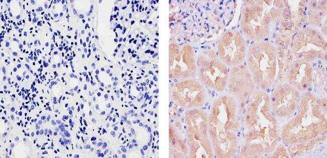
- Immunohistochemistry analysis of Ubiquilin showing staining in the cytoplasm and nucleus of paraffin-embedded mouse kidney tissue (right) compared to a negative control without primary antibody (left). To expose target proteins, antigen retrieval was performed using 10mM sodium citrate (pH 6.0), microwaved for 8-15 min. Following antigen retrieval, tissues were blocked in 3% H2O2-methanol for 15 min at room temperature, washed with ddH2O and PBS, and then probed with a Ubiquilin Rabbit Polyclonal Antibody (Product # PA1-759) diluted in 3% BSA-PBS at a dilution of 1:20 for 1 hour at 37ºC in a humidified chamber. Tissues were washed extensively in PBST and detection was performed using an HRP-conjugated secondary antibody followed by colorimetric detection using a DAB kit. Tissues were counterstained with hematoxylin and dehydrated with ethanol and xylene to prep for mounting.
Supportive validation
- Submitted by
- Invitrogen Antibodies (provider)
- Main image
- Experimental details
- NULL

- Submitted by
- Invitrogen Antibodies (provider)
- Main image
- Experimental details
- NULL
- Submitted by
- Invitrogen Antibodies (provider)
- Main image
- Experimental details
- NULL

- Submitted by
- Invitrogen Antibodies (provider)
- Main image

- Experimental details
- NULL
- Submitted by
- Invitrogen Antibodies (provider)
- Main image
- Experimental details
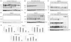
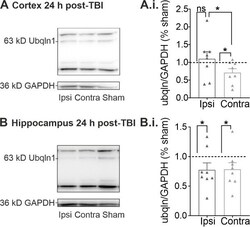
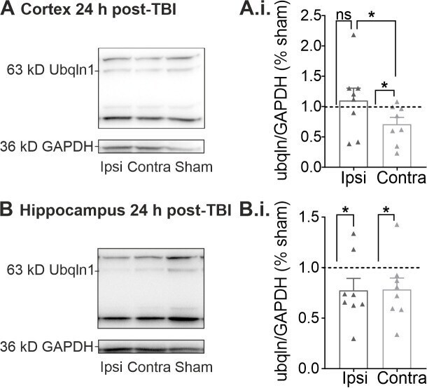
- Quantitative Western Blots of lysates obtained 24 h post-TBI. ( A ) Representative molecular weight bands for ubqln1 (63 kD) and the housekeeper protein GAPDH (36 kD) of lysates obtained from the whole cortex ipsilateral (ipsi), contralateral (contra) and from sham mice 24 h post-TBI. ( A.i. ) The ubqln1 signal per GAPDH signal was normalized to the intensities detected in lysates obtained from sham mice. Note that the expression of ubqln1 was reduced only in the contralateral cortex 24 h post-TBI. ( B ) Representative weight bands for ubqln1 and GAPDH obtained from hippocampus lysates 24 h post-TBI. ( B.i. ) The ubqln1 per-GAPDH expression was lowered in both hippocampal lysates of the contralateral and the ipsilateral hemisphere 24 h post-TBI. The data are represented as mean +- SEM and significant values were detected using a Kruskal-Wallis test with Dunn's multiple comparison, indicated here as * p < 0.05.
- Submitted by
- Invitrogen Antibodies (provider)
- Main image

- Experimental details
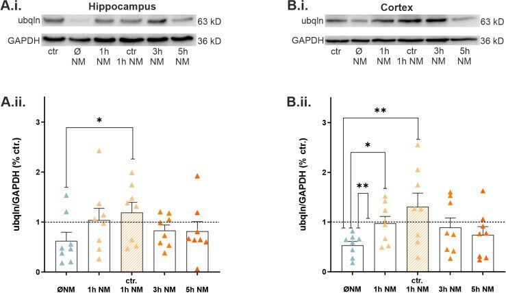
- Expression of hippocampal and cortical levels of ubqln1 following treatment with nialamide (NM) during epilepsy in vitro. ( A.i. ) Representative Western blot weight bands of lysates from hippocampal and cortical ( B.i. ) slices. ( A.ii. ) Scatter plots of hippocampal and cortical ( B.ii. ) ubqln1 (63 kD) expression at various time points of incubation in mACSF or standard ACSF and subsequent treatment with 10 uM NM. The scatter plots summarize data from n = 8 mice, and the data were normalized to the signal of the housekeeping protein GAPDH. The dashed line represents the control group that was kept in standard ACSF and was set to 1. Bar plots indicate mean values +- SEM. Asterisks represent significantly different values received from Kruskal-Wallis tests; they are indicated as * p < 0.05, ** p < 0.01. Note the recovery of ubqln1 expression in the hippocampus during epilepsy in the presence of the MAO inhibitor. We observed even an increase in ubqln1 expression between data from epilepsy conditions in the absence of NM and data from control tissue bathed in standard ACSF, but in the presence of NM (* p = 0.0190). In the cortex, increased expression was disclosed between the epilepsy group in the absence of NM and the control group (** p = 0.0100), as well as the epilepsy group + 1 h NM (* p = 0.0303) and the control + NM (** p = 0.0050), respectively.