 Explore
 Validate
 Learn
Learn Western blot
 ELISA
ELISAAntibody data
- Antibody Data
- Antigen structure
- References [1]
- Comments [0]
- Validations
- Western blot [4]
- Immunocytochemistry [2]
Submit
Validation data
Reference
Comment
Report error
- Product number
- MA5-15670 - Provider product page

- Provider
- Invitrogen Antibodies
- Product name
- RAB10 Monoclonal Antibody (4E2)
- Antibody type
- Monoclonal
- Antigen
- Purifed from natural sources
- Reactivity
- Human, Mouse
- Host
- Mouse
- Isotype
- IgG
- Antibody clone number
- 4E2
- Vial size
- 100 µL
- Concentration
- Conc. Not Determined
- Storage
- Store at 4°C short term. For long term storage, store at -20°C, avoiding freeze/thaw cycles.
Submitted references RAB8, RAB10 and RILPL1 contribute to both LRRK2 kinase-mediated centrosomal cohesion and ciliogenesis deficits.
Lara Ordónez AJ, Fernández B, Fdez E, Romo-Lozano M, Madero-Pérez J, Lobbestael E, Baekelandt V, Aiastui A, López de Munaín A, Melrose HL, Civiero L, Hilfiker S
Human molecular genetics 2019 Nov 1;28(21):3552-3568
Human molecular genetics 2019 Nov 1;28(21):3552-3568
No comments: Submit comment
Supportive validation
- Submitted by
- Invitrogen Antibodies (provider)
- Main image
- Experimental details
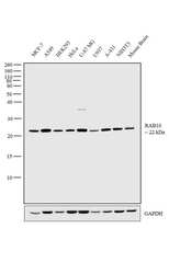
- Western blot analysis was performed on membrane enriched cell extracts (30 µg lysate) of MCF-7 (Lane 1), A549 (Lane 2), HEK293 (Lane 3), HeLa (Lane 4), U-87 MG (Lane 5), U937 (Lane 6), A-431 (Lane 7), NIH3T3 (Lane 8) and tissue extract of Mouse Brain (Lane 9). The blot was probed with Anti-RAB10 Monoclonal Antibody (4E2) (Product # MA5-15670, 1:2000 dilution) and detected by chemiluminescence using Goat anti-Mouse IgG (H+L) Superclonal™ Secondary Antibody, HRP conjugate (Product # A28177, 0.25 µg/ml, 1:4000 dilution). A 22 kDa band corresponding to RAB10 was observed across all the cell lines and tissue tested.

- Submitted by
- Invitrogen Antibodies (provider)
- Main image

- Experimental details
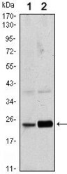
- Western blot analysis of Rab10 using Rab10 monoclonal antibody (Product # MA5-15670) in HeLa (1) and NIH/3T3 (2) cell lysate.

- Submitted by
- Invitrogen Antibodies (provider)
- Main image
- Experimental details
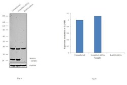
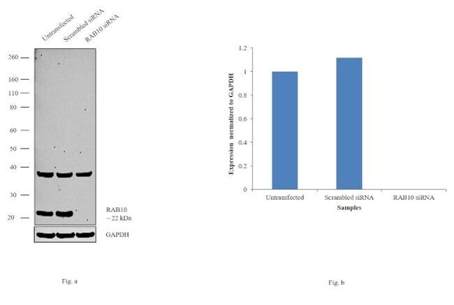
- Knockdown of RAB10 was achieved by transfecting HeLa cells with RAB10 specific siRNAs (Silencer® select Product # s21391, s21392). Western blot analysis (Fig. a) was performed using membrane enriched extracts from the RAB10 knockdown cells (lane 3), non-specific scrambled siRNA transfected cells (lane 2) and untransfected cells (lane 1). The blot was probed with RAB10 Monoclonal Antibody (Product # MA5-15670, 1:2000 dilution) and Goat anti-Mouse IgG (H+L) Superclonal™ Secondary Antibody, HRP conjugate (Product # A28177, 0.25µg/ml, 1:4000 dilution). Densitometric analysis of this western blot is shown in histogram (Fig. b). Decrease in signal upon siRNA mediated knock down confirms that antibody is specific to RAB10.
- Submitted by
- Invitrogen Antibodies (provider)
- Main image
- Experimental details
- Western blot analysis of Rab10 using Rab10 monoclonal antibody (Product # MA5-15670) in HeLa (1) and NIH/3T3 (2) cell lysate.

Supportive validation
- Submitted by
- Invitrogen Antibodies (provider)
- Main image
- Experimental details
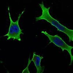
- Immunofluorescence analysis of LOVO cells using Rab10 monoclonal antibody (Product # MA5-15670) (Green). Blue: DRAQ5 fluorescent DNA dye.

- Submitted by
- Invitrogen Antibodies (provider)
- Main image
- Experimental details
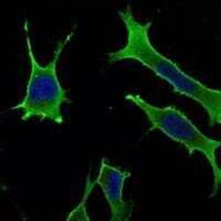
- Immunofluorescence analysis of LOVO cells using Rab10 monoclonal antibody (Product # MA5-15670) (Green). Blue: DRAQ5 fluorescent DNA dye.
