Explore
 Validate
 Learn
Learn Western blot
 Immunocytochemistry
Antibody data
- Antibody Data
- Antigen structure
- References [7]
- Comments [0]
- Validations
- Western blot [1]
- Immunohistochemistry [1]
- Flow cytometry [6]
Submit
Validation data
Reference
Comment
Report error
- Product number
- NB300-524 - Provider product page

- Provider
- Novus Biologicals
- Proper citation
- Novus Cat#NB300-524, RRID:AB_10001871
- Product name
- O-GlcNAc Antibody (RL2)
- Antibody type
- Monoclonal
- Antigen
- Pore complex-lamina fraction purified from rat liver nuclear envelopes.
- Reactivity
- Human, Mouse, Rat
- Host
- Mouse
- Isotype
- IgG
Submitted references Interplay Between Phosphorylation and O-GlcNAcylation of Sarcomeric Proteins in Ischemic Heart Failure.
O-GlcNAcylation is required for mutant KRAS-induced lung tumorigenesis.
Glucose and glutamine fuel protein O-GlcNAcylation to control T cell self-renewal and malignancy.
Removal of O-GlcNAcylation is important for pig preimplantation development.
O-GlcNAcylation of the human epidermal growth factor receptor.
O-GlcNAc transferase integrates metabolic pathways to regulate the stability of c-MYC in human prostate cancer cells.
O-GlcNAcylation of kinases.
Mercier T, Bouvet M, Dubois-Deruy E, Dechaumes A, Beseme O, Richard V, Mulder P, Pinet F
Frontiers in endocrinology 2018;9:598
Frontiers in endocrinology 2018;9:598
O-GlcNAcylation is required for mutant KRAS-induced lung tumorigenesis.
Taparra K, Wang H, Malek R, Lafargue A, Barbhuiya MA, Wang X, Simons BW, Ballew M, Nugent K, Groves J, Williams RD, Shiraishi T, Verdone J, Yildirir G, Henry R, Zhang B, Wong J, Wang KK, Nelkin BD, Pienta KJ, Felsher D, Zachara NE, Tran PT
The Journal of clinical investigation 2018 Nov 1;128(11):4924-4937
The Journal of clinical investigation 2018 Nov 1;128(11):4924-4937
Glucose and glutamine fuel protein O-GlcNAcylation to control T cell self-renewal and malignancy.
Swamy M, Pathak S, Grzes KM, Damerow S, Sinclair LV, van Aalten DM, Cantrell DA
Nature immunology 2016 Jun;17(6):712-20
Nature immunology 2016 Jun;17(6):712-20
Removal of O-GlcNAcylation is important for pig preimplantation development.
Shibutani M, Mori T, Miyano T, Miyake M
The Journal of reproduction and development 2015;61(4):341-50
The Journal of reproduction and development 2015;61(4):341-50
O-GlcNAcylation of the human epidermal growth factor receptor.
Stateva SR, Villalobo A
Organic & biomolecular chemistry 2015 Aug 14;13(30):8196-204
Organic & biomolecular chemistry 2015 Aug 14;13(30):8196-204
O-GlcNAc transferase integrates metabolic pathways to regulate the stability of c-MYC in human prostate cancer cells.
Itkonen HM, Minner S, Guldvik IJ, Sandmann MJ, Tsourlakis MC, Berge V, Svindland A, Schlomm T, Mills IG
Cancer research 2013 Aug 15;73(16):5277-87
Cancer research 2013 Aug 15;73(16):5277-87
O-GlcNAcylation of kinases.
Dias WB, Cheung WD, Hart GW
Biochemical and biophysical research communications 2012 Jun 1;422(2):224-8
Biochemical and biophysical research communications 2012 Jun 1;422(2):224-8
No comments: Submit comment
Supportive validation
- Submitted by
- Novus Biologicals (provider)
- Main image

- Experimental details
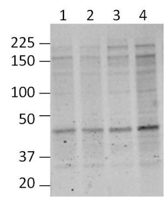
- Western Blot: O-GlcNAc Antibody (RL2) [NB300-524] - Analysis of mouse cortical brain lysates using O-Linked N-Acetylglucosamine Monoclonal Antibody. Blots containing cortical extracts from 4 individual C57BL/6 mice (Lanes 1-4) were blocked with 5% milk in TBST, and probed with MA1-072 at 1:1000, followed by a fluorophore-conjugated goat anti-mouse IgG secondary antibody. Data courtesy of the Innovators Program.
Supportive validation
- Submitted by
- Novus Biologicals (provider)
- Main image

- Experimental details
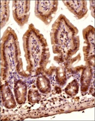
- Immunohistochemistry-Paraffin: O-GlcNAc Antibody (RL2) [NB300-524] - Analysis of a FFPE tissue section of the mouse colon using 1:200 dilution of O-GlcNAc [RL2] antibody (NB300-524). The signal was developed using HRP-DAB method which followed counterstaining of the cells with hematoxylin.
Supportive validation
- Submitted by
- Novus Biologicals (provider)
- Main image
- Experimental details
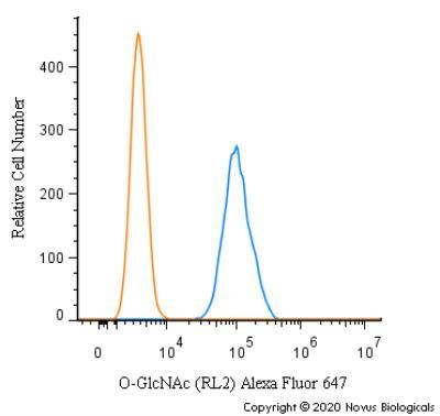
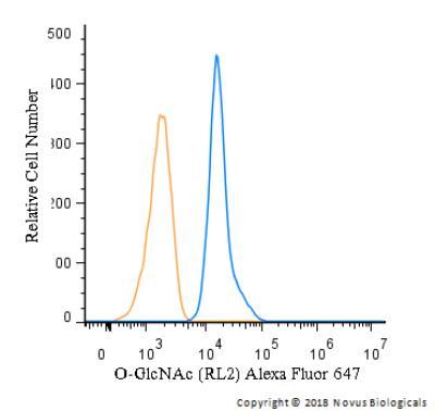
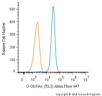
- Flow Cytometry: O-GlcNAc Antibody (RL2) [NB300-524] - Analysis using Alexa Fluor (R) 647 conjugate of NB300-524. An intracellular stain was performed on Jurkat cells with O-GlcNAc antibody (RL2) NB300-524 (blue) and a matched isotype control NBP2-27287 (orange). Cells were fixed with 4% PFA and then permeablized with 0.1% saponin. 1 ug of antibody was added to 100 uL of staining buffer and cells were incubated for 30 minutes at room temperature. Both antibodies were conjugated to Alexa Fluor 647.

- Submitted by
- Novus Biologicals (provider)
- Main image

- Experimental details
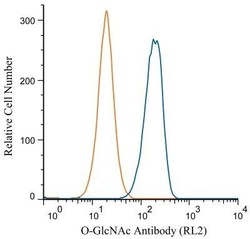
- Flow Cytometry: O-GlcNAc Antibody (RL2) [NB300-524] - An intracellular stain was performed on U-937 cells with O-GlcNAc antibody (RL2) NB300-524AF647 (blue) and a matched isotype control. Cells were fixed with 4% PFA and then permeablized with 0.1% saponin. Cells were incubated in an antibody dilution of 2.5 ug/mL for 30 minutes at room temperature . Both antibodies were conjugated to Alexa Fluor 647.
- Submitted by
- Novus Biologicals (provider)
- Main image
- Experimental details
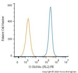
- Flow Cytometry: O-GlcNAc Antibody (RL2) [NB300-524] - An intracellular stain was performed on Jurkat cells with O-GlcNAc antibody (RL2) NB300-524PE (blue) and a matched isotype control. Cells were fixed with 4% PFA and then permeablized with 0.1% saponin. Cells were incubated in an antibody dilution of 2.5 ug/mL for 30 minutes at room temperature . Both antibodies were conjugated to Phycoerythrin.
- Submitted by
- Novus Biologicals (provider)
- Main image
- Experimental details
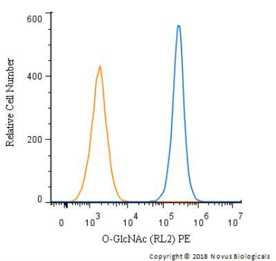
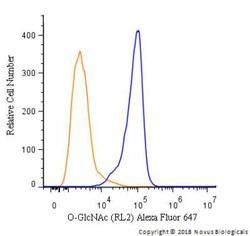
- Flow Cytometry: O-GlcNAc Antibody (RL2) [NB300-524] - An intracellular stain was performed on SK-MEL-28 cells with O-GlcNAc antibody (RL2) NB300-524AF647 (blue) and a matched isotype control. Cells were fixed with 4% PFA and then permeablized with 0.1% saponin. Cells were incubated in an antibody dilution of 2.5 ug/mL for 30 minutes at room temperature . Both antibodies were conjugated to Alexa Fluor 647.
- Submitted by
- Novus Biologicals (provider)
- Main image
- Experimental details
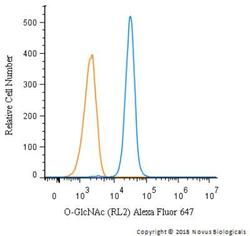
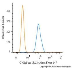
- Flow Cytometry: O-GlcNAc Antibody (RL2) [NB300-524] - An intracellular stain was performed on HeLa cells with O-GlcNAc Antibody [RL2] Antibody NB300-524AF647 (blue) and a matched isotype control (orange). Cells were fixed with 4% PFA and then permeabilized with 0.1% saponin. Cells were incubated in an antibody dilution of 2.5 ug/mL for 30 minutes at room temperature. Both antibodies were conjugated to Alexa Fluor 647.

- Submitted by
- Novus Biologicals (provider)
- Main image
- Experimental details
- Flow Cytometry: O-GlcNAc Antibody (RL2) [NB300-524] - An intracellular stain was performed on RH30 cells with O-GlcNAc [RL2] Antibody NB300-524AF647 (blue) and a matched isotype control (orange). Cells were fixed with 4% PFA and then permeabilized with 0.1% saponin. Cells were incubated in an antibody dilution of 2.5 ug/mL for 30 minutes at room temperature. Both antibodies were conjugated to Alexa Fluor 647.