 Explore
 Validate
 Learn
Learn Western blot
 ELISA
ELISAAntibody data
- Antibody Data
- Antigen structure
- References [0]
- Comments [0]
- Validations
- Western blot [1]
Submit
Validation data
Reference
Comment
Report error
- Product number
- LS-C154009 - Provider product page

- Provider
- LSBio
- Product name
- GDF15 Antibody (clone 23B3.D2.H5) LS-C154009
- Antibody type
- Monoclonal
- Description
- Protein A affinity chromatography
- Reactivity
- Human
- Host
- Mouse
- Isotype
- IgG
- Antibody clone number
- 23B3.D2.H5
- Storage
- Short term: store at 4°C. Long term: aliquot and store at -20°C. Avoid freeze-thaw cycles.
No comments: Submit comment
Supportive validation
- Submitted by
- LSBio (provider)
- Enhanced method
- Genetic validation
- Main image
- Experimental details
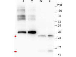
- Anti-NAG-1 Monoclonal Antibody - Western Blot. Western blot of anti-NAG-1 monoclonal antibody. The blot shows detection of recombinant NAG-1 protein present in Pichia pastoris whole cell lysates: lane 1 - yeast cell lysate expressing NAG-1 H variant with SUMO expression tag at 36 kD; lane 2 - yeast cell lysate expressing NAG-1 D variant with SUMO expression tag at 36 kD; lane 3 - yeast cell lysate expressing NAG-1 H variant; and lane 4 - yeast cell lysate expressing NAG-1 D variant. Recombinant NAG-1 proteins without SUMO correspond to monomer (15 kD) and dimer (30 kD) bands as indicated by the arrowheads. All lysates were run under reducing conditions. Primary antibody was used at a 1:1000 dilution in TBS containing 1% BSA and 0.2% Tween, and reacted overnight at 4C. For detection, a 1:40000 dilution of peroxidase conjugated Gt-a-Mouse IgG secondary antibody (LS-C60680) was used in Blocking Buffer for Fluorescent Western Blot (MB-070) for 30 min at room temperature. Molecular weight estimation was made by comparison to prestained MW markers. Image was captured using the BioRad Versadoc 4000MP Imaging System.
