Explore
 Validate
 Learn
Learn Western blot
Antibody data
- Antibody Data
- Antigen structure
- References [2]
- Comments [0]
- Validations
- Western blot [3]
- Immunocytochemistry [2]
- Immunohistochemistry [1]
Submit
Validation data
Reference
Comment
Report error
- Product number
- GTX101703 - Provider product page

- Provider
- GeneTex
- Proper citation
- GeneTex Cat#GTX101703, RRID:AB_1951203
- Product name
- PGD antibody [N1N3]
- Antibody type
- Polyclonal
- Reactivity
- Human, Mouse
- Host
- Rabbit
Submitted references A clinical drug library screen identifies clobetasol propionate as an NRF2 inhibitor with potential therapeutic efficacy in KEAP1 mutant lung cancer.
Quiescent fibroblasts exhibit high metabolic activity.
Choi EJ, Jung BJ, Lee SH, Yoo HS, Shin EA, Ko HJ, Chang S, Kim SY, Jeon SM
Oncogene 2017 Sep 14;36(37):5285-5295
Oncogene 2017 Sep 14;36(37):5285-5295
Quiescent fibroblasts exhibit high metabolic activity.
Lemons JM, Feng XJ, Bennett BD, Legesse-Miller A, Johnson EL, Raitman I, Pollina EA, Rabitz HA, Rabinowitz JD, Coller HA
PLoS biology 2010 Oct 19;8(10):e1000514
PLoS biology 2010 Oct 19;8(10):e1000514
No comments: Submit comment
Supportive validation
- Submitted by
- GeneTex (provider)
- Main image
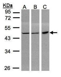
- Experimental details
- Sample(30 ?g whole cell lysate)A:293TB:MOLT4 (GTX27912)C:Raji (GTX27908)10% SDS PAGEGTX101703 diluted at 1:1000The HRP-conjugated anti-rabbit IgG antibody (GTX213110-01) was used to detect the primary antibody.
- Submitted by
- GeneTex (provider)
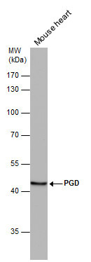
- Main image
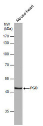
- Experimental details
- PGD antibody [N1N3] detects PGD protein by western blot analysis. Mouse tissue extracts (50 ?g) was separated by 10% SDS-PAGE, and the membrane was blotted with PGD antibody [N1N3] (GTX101703) diluted at 1:1000. The HRP-conjugated anti-rabbit IgG antibody (GTX213110-01) was used to detect the primary antibody.
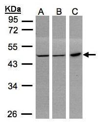
- Submitted by
- GeneTex (provider)
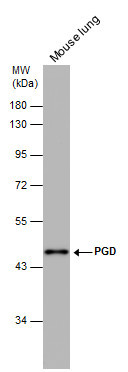
- Main image
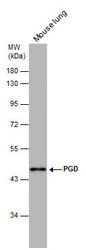
- Experimental details
- PGD antibody [N1N3] detects PGD protein by western blot analysis. Mouse tissue extracts (50 ?g) was separated by 10% SDS-PAGE, and the membrane was blotted with PGD antibody [N1N3] (GTX101703) diluted at 1:500. The HRP-conjugated anti-rabbit IgG antibody (GTX213110-01) was used to detect the primary antibody.
Supportive validation
- Submitted by
- GeneTex (provider)
- Main image

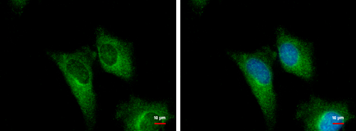
- Experimental details
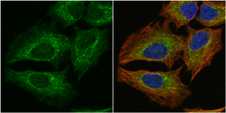
- PGD antibody [N1N3] detects PGD protein at cytoplasm by immunofluorescent analysis.Sample: HeLa cells were fixed in ice-cold MeOH for 5 min.Green: PGD protein stained by PGD antibody [N1N3] (GTX101703) diluted at 1:500.Blue: Hoechst 33342 staining.
- Submitted by
- GeneTex (provider)
- Main image
- Experimental details
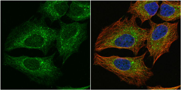
- PGD antibody [N1N3] detects PGD protein at cytoplasm by immunofluorescent analysis.Sample: HeLa cells were fixed in 4% paraformaldehyde at RT for 15 min.Green: PGD protein stained by PGD antibody [N1N3] (GTX101703) diluted at 1:1000.Red: alpha Tubulin, a cytoskeleton marker, stained by alpha Tubulin antibody [GT114] (GTX628802) diluted at 1:1000.Blue: Hoechst 33342 staining.
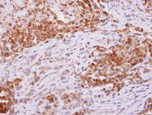
Supportive validation
- Submitted by
- GeneTex (provider)
- Main image

- Experimental details
- PGD antibody [N1N3] detects PGD protein at cytoplasm on A549 xenograft by immunohistochemical analysis. Sample: Paraffin-embedded A549 xenograft. PGD antibody [N1N3] (GTX101703) dilution: 1:500.