Explore
 Validate
 Learn
Learn Western blot
Antibody data
- Antibody Data
- Antigen structure
- References [1]
- Comments [0]
- Validations
- Western blot [5]
- Immunocytochemistry [2]
- Immunohistochemistry [1]
Submit
Validation data
Reference
Comment
Report error
- Product number
- PA5-21376 - Provider product page

- Provider
- Invitrogen Antibodies
- Product name
- PGD Polyclonal Antibody
- Antibody type
- Polyclonal
- Antigen
- Recombinant protein fragment
- Description
- Recommended positive controls: 293T, Molt-4, Raji, Mouse heart, Mouse lung.
- Concentration
- 1 mg/mL
Submitted references A feedback loop between the androgen receptor and 6-phosphogluoconate dehydrogenase (6PGD) drives prostate cancer growth.
Gillis JL, Hinneh JA, Ryan NK, Irani S, Moldovan M, Quek LE, Shrestha RK, Hanson AR, Xie J, Hoy AJ, Holst J, Centenera MM, Mills IG, Lynn DJ, Selth LA, Butler LM
eLife 2021 Aug 12;10
eLife 2021 Aug 12;10
No comments: Submit comment
Supportive validation
- Submitted by
- Invitrogen Antibodies (provider)
- Main image
- Experimental details
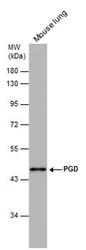
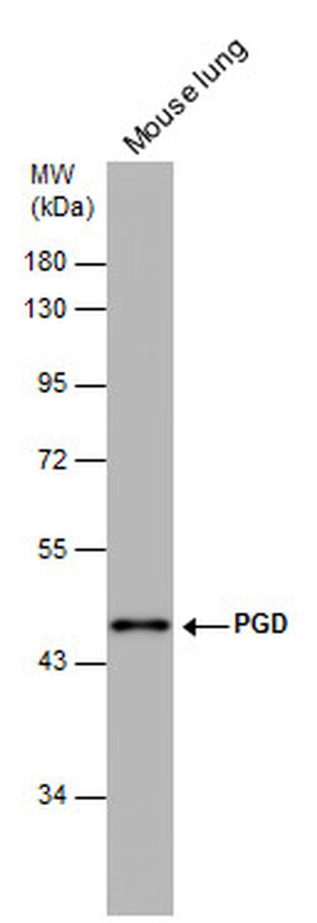
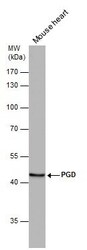
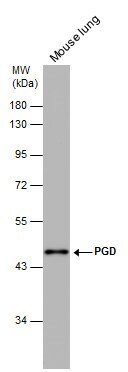
- Western blot analysis of PGD in Mouse tissue extracts (50 µg). Samples was separated by 10% SDS-PAGE and the membrane was probed with PGD Polyclonal antibody (Product # PA5-21376) at a dilution of 1:500.
- Submitted by
- Invitrogen Antibodies (provider)
- Main image
- Experimental details
- Western blot analysis of PGD in Mouse tissue extracts (50 µg). Samples was separated by 10% SDS-PAGE and the membrane was probed with PGD Polyclonal antibody (Product # PA5-21376) at a dilution of 1:1000.

- Submitted by
- Invitrogen Antibodies (provider)
- Main image
- Experimental details
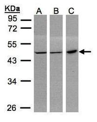
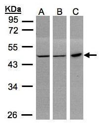
- Western Blot using PGD Polyclonal Antibody (Product # PA5-21376). Sample (30 µg whole cell lysate). A: 293T. B: MOLT4. C: Raji. 10% SDS PAGE. PGD Polyclonal Antibody (Product # PA5-21376) diluted at 1:1,000. The HRP-conjugated anti-rabbit IgG antibody was used to detect the primary antibody.
- Submitted by
- Invitrogen Antibodies (provider)
- Main image
- Experimental details
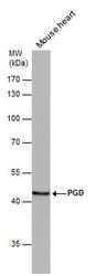
- PGD Polyclonal Antibody detects PGD protein by western blot analysis. Mouse tissue extracts (50 µg) was separated by 10% SDS-PAGE, and the membrane was blotted with PGD Polyclonal Antibody (Product # PA5-21376) diluted at 1:1,000. The HRP-conjugated anti-rabbit IgG antibody was used to detect the primary antibody.

- Submitted by
- Invitrogen Antibodies (provider)
- Main image

- Experimental details
- PGD Polyclonal Antibody detects PGD protein by western blot analysis. Mouse tissue extracts (50 µg) was separated by 10% SDS-PAGE, and the membrane was blotted with PGD Polyclonal Antibody (Product # PA5-21376) diluted at 1:500. The HRP-conjugated anti-rabbit IgG antibody was used to detect the primary antibody.
Supportive validation
- Submitted by
- Invitrogen Antibodies (provider)
- Main image
- Experimental details
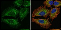
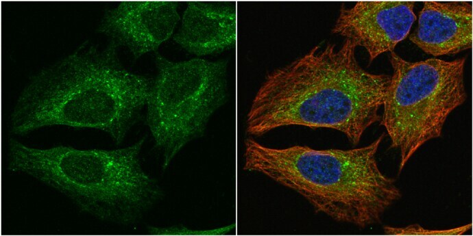
- Immunocytochemistry-Immunofluorescence analysis of PGD was performed in HeLa cells fixed in 4% paraformaldehyde at RT for 15 min. Green: PGD Polyclonal Antibody (Product # PA5-21376) diluted at 1:1000. Red: alpha Tubulin, a cytoskeleton marker. Blue: Hoechst 33342 staining.
- Submitted by
- Invitrogen Antibodies (provider)
- Main image
- Experimental details
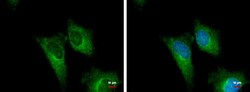
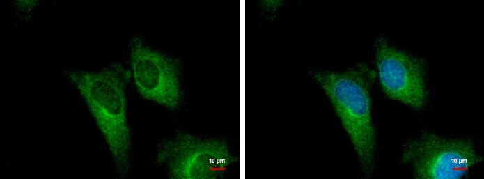
- PGD Polyclonal Antibody detects PGD protein at cytoplasm by immunofluorescent analysis. Sample: HeLa cells were fixed in ice-cold MeOH for 5 min. Green: PGD protein stained by PGD Polyclonal Antibody (Product # PA5-21376) diluted at 1:500. Blue: Hoechst 33342 staining.
Supportive validation
- Submitted by
- Invitrogen Antibodies (provider)
- Main image

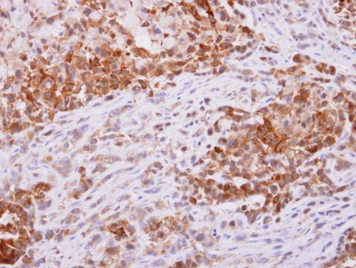
- Experimental details
- PGD Polyclonal Antibody detects PGD protein at cytoplasm on A549 xenograft by immunohistochemical analysis. Sample: Paraffin-embedded A549 xenograft. PGD Polyclonal Antibody (Product # PA5-21376) dilution: 1:500. Antigen Retrieval: EDTA based buffer, pH 8.0, 15 min.