Explore
 Validate
 Learn
Learn Western blot
Antibody data
- Antibody Data
- Antigen structure
- References [1]
- Comments [0]
- Validations
- Western blot [2]
- Immunohistochemistry [6]
Submit
Validation data
Reference
Comment
Report error
- Product number
- TA319485 - Provider product page

- Provider
- OriGene
- Product name
- Rabbit polyclonal anti-AHSP antibody
- Antibody type
- Polyclonal
- Description
- Rabbit polyclonal anti-AHSP antibody
- Host
- Rabbit
- Conjugate
- Unconjugated
- Epitope
- AHSP
- Isotype
- IgG
- Antibody clone number
- NULL
- Vial size
- 100 µl
- Concentration
- 86mg/ml
Submitted references Hsp90 chaperones hemoglobin maturation in erythroid and nonerythroid cells.
Ghosh A, Garee G, Sweeny EA, Nakamura Y, Stuehr DJ
Proceedings of the National Academy of Sciences of the United States of America 2018 Feb 6;115(6):E1117-E1126
Proceedings of the National Academy of Sciences of the United States of America 2018 Feb 6;115(6):E1117-E1126
No comments: Submit comment
Supportive validation
- Submitted by
- OriGene (provider)
- Main image

- Experimental details
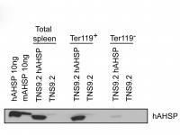
- Lane 1: Recombinant hAHSP. Lane 2: Recombinant mAHSP. Lane 3: mice Spleen cells transfected with TNS9.2-hAHSP. Lane 4: mice Spleen cells transfected with TNS9.2 control vector. Lane 5: mice Spleen cells transfected with TNS9.2-hAHSP fractionated by MACS using Ter119+ microbeads. Lane 6: mice Spleen cells transfected with TNS9.2 control vector fractionated by Ter119+. Lane 7: mice Spleen cells transfected with TNS9.2-hAHSP fractionated by Ter119-. Lane 8: Spleen cells from mice transduced with TNS9.2 control vector fractionated by Ter119-. Load: 10ng per lane. Anti-AHSP: 1:1,000; Secondary: HRP Streptavidin at 1:40,000.
- Validation comment
- WB
- Submitted by
- OriGene (provider)
- Main image
- Experimental details
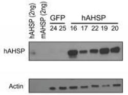
- WB of Rabbit anti-AHSP antibody. Lane 1: Recombinant hAHSP (2 ng). Lane 2: Recombinant mAHSP (2 ng). Lane 3: RBC Lysates Mouse #24 - GFP. Lane 4: RBC Lysates Mouse #25 - GFP. Lane 5: RBC Lysates Mouse #16 - hAHSP. Lane 6: RBC Lysates Mouse #17 - hAHSP. Lane 7: RBC Lysates Mouse #22 - hAHSP. Lane 8: RBC Lysates Mouse #19 - hAHSP. Lane 9: RBC Lysates Mouse #20 - hAHSP. Primary antibody: hAHSP antibody, Beta-Actin antibody at 1:1,000. Secondary antibody:1:40,000.
- Validation comment
- WB

Supportive validation
- Submitted by
- OriGene (provider)
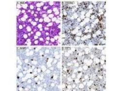
- Main image

- Experimental details
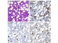
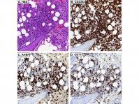
- IHC of Rabbit anti-AHSP antibody. Tissue: A. Normal bone marrow, H&E. B. CD235a stains both nucleated EPs and mature, anucleate RBCs. C. AHSP stains nucleated EPs, but not mature, anucleate RBCs. D. CD71 stains nucleated EPs, but not mature, anucleate RBCs. Primary antibody: AHSP antibody at 1:8,000 for overnight at 4°C Secondary antibody: anti-rabbit secondary at (1:10,000 for 45 min at RT).
- Validation comment
- IHC
- Submitted by
- OriGene (provider)
- Main image

- Experimental details
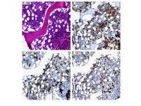
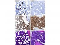
- IHC of Rabbit anti-AHSP antibody. Tissue: A. Acute erythroleukemia, H&E. B. CD235a stains erythroid blasts and mature, anucleate RBCs. C. AHSP stains erythroid blasts. D. CD71 stains erythroid blasts. Primary antibody: AHSP antibody at 1:8,000 for overnight at 4°C Secondary antibody: anti-rabbit secondary at (1:10,000 for 45 min at RT)
- Validation comment
- IHC
- Submitted by
- OriGene (provider)
- Main image

- Experimental details
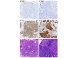
- IHC of Rabbit anti-AHSP antibody. Tissue: AHSP stains residual EPs and not myeloid blasts in acute myeloid leukemia with minimal differentiation (A), whereas CD71 stains both myeloid blasts and EPs (B). AHSP does not stain myeloid blasts in acute myelomonocytic leukemia (D), whereas CD71 does (E). C and F are corresponding H&Es, respectively. Primary antibody: AHSP antibody at 1:8,000 for overnight at 4°C Secondary antibody: anti-rabbit secondary at (1:10,000 for 45 min at RT).
- Validation comment
- IHC
- Submitted by
- OriGene (provider)
- Main image

- Experimental details
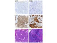
- IHC of Rabbit anti-AHSP antibody. Tissue: A. Primary myelofibrosis, H&E. B. CD235a stains both nucleated EPs and mature, anucleate RBCs. AHSP C. and CD71 D. variably stain megakaryocytes and also stain nucleated EPs. Primary antibody: AHSP antibody at 1:8,000 for overnight at 4°C Secondary antibody: anti-rabbit secondary at (1:10,000 for 45 min at RT).
- Validation comment
- IHC

- Submitted by
- OriGene (provider)
- Main image
- Experimental details
- IHC of Rabbit anti-AHSP antibody. Tissue: AHSP A. stains residual EPs and not lymphoma cells in DLBCL, whereas CD71 B. stains both lymphoma cells and EPs. C. Corresponding H&E. AHSP D. does not metastatic carcinoma, whereas CD71 E. does. F. Corresponding H&E. Primary antibody: AHSP antibody at 1:8,000 for overnight at 4°C Secondary antibody: anti-rabbit secondary at (1:10,000 for 45 min at RT).
- Validation comment
- IHC
- Submitted by
- OriGene (provider)
- Main image
- Experimental details
- IHC of Rabbit anti-AHSP antibody. Tissue: Giant pronormoblasts are evident in parvoviral infection (H&E, A). B. CD235a does not stain giant pronormoblasts. AHSP C. and CD71 D. stain giant pronormoblasts. Primary antibody: AHSP antibody at 1:8,000 for overnight at 4°C Secondary antibody: anti-rabbit secondary at (1:10,000 for 45 min at RT) .
- Validation comment
- IHC