Explore
 Validate
 Learn
Learn Western blot
Antibody data
- Antibody Data
- Antigen structure
- References [0]
- Comments [0]
- Validations
- Western blot [2]
- Immunohistochemistry [1]
Submit
Validation data
Reference
Comment
Report error
- Product number
- PAB9961 - Provider product page

- Provider
- Abnova Corporation
- Proper citation
- Abnova Corporation Cat#PAB9961, RRID:AB_1710200
- Product name
- MYL6 (phospho S19/20) polyclonal antibody
- Antibody type
- Polyclonal
- Description
- Rabbit polyclonal antibody raised against synthetic phosphopeptide of MYL6.
- Storage
- Store at 4°C. For long term storage store at -20°C.Aliquot to avoid repeated freezing and thawing.
No comments: Submit comment
Supportive validation
- Submitted by
- Abnova Corporation (provider)
- Main image
- Experimental details
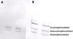
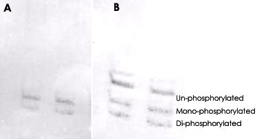
- MYL6 (phospho S19/20) polyclonal antibody (Cat # PAB9961) was used at a 1 : 1000 dilution to detect myosin light chain by Western blot on NIH/3T3 cell lysates.A standard urea/glycerol gel without SDS was used to separate phospho forms of regulatory light chain according to mass to charge ratios.In Panel A, reactivity of MYL6 (phospho S19/20) polyclonal antibody (Cat # PAB9961) is shown.In Panel B, reactivity of commercially available pan reactive antibody that detects both unphosphorylated and phosphorylated forms of regulatory light chain is shown.The phosphospecific antibody detects both monophosphorylated (pSer20 Mono-P-RLC) and diphosphorylated (pThr19-pSer20 Di-P-RLC) regulatory light chain.Personal communication. J. Stull. UT Southwestern Medical Center.
- Submitted by
- Abnova Corporation (provider)
- Main image
- Experimental details
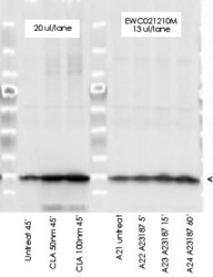
- MYL6 (phospho S19/20) polyclonal antibody (Cat # PAB9961) was used at a 1 : 5000 dilution to detect myosin light chain by Western blot.Either 13 or 20 uL of a mouse cardiac myocyte lysate was loaded on a 4-20% Criterion gel for SDS-PAGE. Samples were either mock-treated or CLA-treated, as indicated. After washing, a 1 : 5,000 dilution of HRP conjugated Gt-a-Rabbit IgG preceded color development using Amersham's substrate system.Other detection methods will yield similar results.Data courtesy of the Alliance for Cellular Signaling (http://www.signaling-gateway.org).

Supportive validation
- Submitted by
- Abnova Corporation (provider)
- Main image

- Experimental details
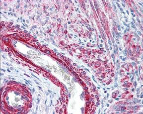
- Immunohistochemical staining with MYL6 (phospho S19/20) polyclonal antibody (Cat # PAB9961) was used at 2.5 ug/mL to detect signal in a variety of tissues including multi-human, multi-brain and multi-cancer slides.This image shows strong staining of both vascular and myometrial smooth muscle cells of the uterus.Tissue was formalin-fixed and paraffin embedded.The image shows localization of the antibody as the precipitated red signal, with a hematoxylin purple nuclear counterstain. Personal Communication, Tina Roush, LifeSpanBiosciences, Seattle, WA.
- Validation comment
- Immunohistochemistry (Formalin/PFA-fixed paraffin-embedded sections)