 Explore
 Validate
 Learn
Learn Western blot
Antibody data
- Antibody Data
- Antigen structure
- References [0]
- Comments [0]
- Validations
- Western blot [2]
- Immunohistochemistry [1]
- Flow cytometry [1]
Submit
Validation data
Reference
Comment
Report error
- Product number
- AP17856PU-N - Provider product page

- Provider
- Acris Antibodies GmbH
- Proper citation
- Acris Antibodies GmbH Cat#AP17856PU-N, RRID:AB_10654518
- Product name
- anti ZW10 interactor / ZWINT (Center)
- Antibody type
- Polyclonal
- Antigen
- KLH conjugated synthetic peptide between 59-88 selected from the Center region of Human ZWINT
- Reactivity
- Human, Mouse
- Host
- Rabbit
- Vial size
- 0.4 ml
- Concentration
- 2.0 mg/ml
No comments: Submit comment
Supportive validation
- Submitted by
- Acris Antibodies GmbH (provider)
- Main image
- Experimental details
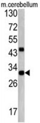
- Western blot analysis: AP17856PU-N ZWINT Antibody staining of Mouse cerebellum tissue lysates (35ug/lane). ZWINT (arrow) was detected using the purified Pab.

- Submitted by
- Acris Antibodies GmbH (provider)
- Main image

- Experimental details
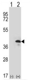
- Western blot analysis: AP17856PU-N ZWINT Antibody staining of 293 cell lysates (2 µg/lane) either nontransfected (Lane 1) or transiently transfected (Lane 2) with the ZWINT gene.
Supportive validation
- Submitted by
- Acris Antibodies GmbH (provider)
- Main image

- Experimental details
- Immunohitochemistry: AP17856PU-N ZWINT Antibody staining of Formalin-Fixed, Paraffin-Embedded Human Brain Tissue. This ZWINT antibody (Center) was peroxidase-conjugated to the secondary antibody, followed by DAB staining. This data demonstrates the use of this antibody for immunohistochemistry. Clinical relevance has not been evaluated.

Supportive validation
- Submitted by
- Acris Antibodies GmbH (provider)
- Main image
- Experimental details
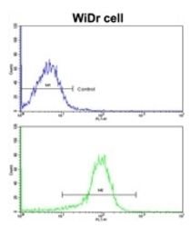
- Flow Cytometry: Analysis of widr cells using AP17856PU-N ZWINT Antibody (Center)(bottom histogram) compared to a Negative control cell (Top histogram). FITC-conjugated Goat-anti-Rabbit secondary antibodies were used for the analysis.
