 Explore
 Validate
 Learn
Learn Western blot
Antibody data
- Antibody Data
- Antigen structure
- References [2]
- Comments [0]
- Validations
- Western blot [1]
- Immunocytochemistry [1]
- Immunohistochemistry [2]
Submit
Validation data
Reference
Comment
Report error
- Product number
- MA3-701 - Provider product page

- Provider
- Invitrogen Antibodies
- Product name
- CRMP5 Monoclonal Antibody (CR-1)
- Antibody type
- Monoclonal
- Antigen
- Purifed from natural sources
- Description
- MA3-701 detects human, bovine, rat and C-terminal region of CRMP5. This antibody has not been shown to be reactive in mouse samples.
- Antibody clone number
- CR-1
- Concentration
- Conc. Not Determined
Submitted references Paraneoplastic autoimmune optic neuritis with retinitis defined by CRMP-5-IgG.
CRMP-5 neuronal autoantibody: marker of lung cancer and thymoma-related autoimmunity.
Cross SA, Salomao DR, Parisi JE, Kryzer TJ, Bradley EA, Mines JA, Lam BL, Lennon VA
Annals of neurology 2003 Jul;54(1):38-50
Annals of neurology 2003 Jul;54(1):38-50
CRMP-5 neuronal autoantibody: marker of lung cancer and thymoma-related autoimmunity.
Yu Z, Kryzer TJ, Griesmann GE, Kim K, Benarroch EE, Lennon VA
Annals of neurology 2001 Feb;49(2):146-54
Annals of neurology 2001 Feb;49(2):146-54
No comments: Submit comment
Supportive validation
- Submitted by
- Invitrogen Antibodies (provider)
- Main image

- Experimental details
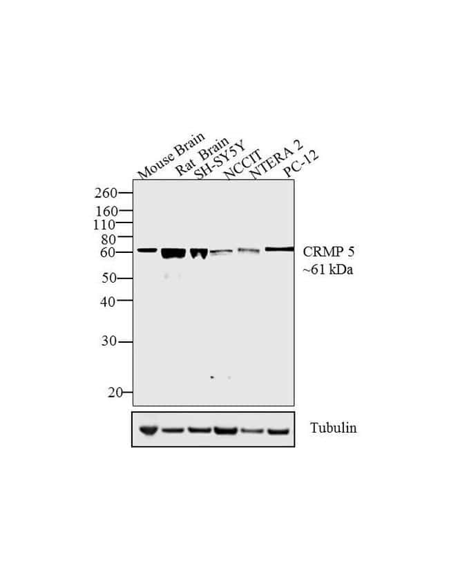
- Western blot analysis was performed on tissue, whole cell extracts (30 µg lysate) of Mouse Brain (Lane 1), Rat Brain (lane 2), SH-SY5Y (lane 3), NCCIT (Lane 4), NTERA 2 (Lane 5), and PC 12 (lane 6). The blots were probed with Anti-CRMP5 Rat Monoclonal Antibody (Product # MA3-701, 1 in 1000 dilution) and detected by chemiluminescence Goat Anti-Rat IgG (H+L) Secondary Antibody, HRP conjugate (Product # 62-9520, 1:1000 dilution). A 61 kDa band corresponding to CRMP5 was observed across cell lines tested. Known quantity of protein samples were electrophoresed using Novex® NuPAGE® 10 % Bis-Tris gel (Product # NP0301BOX), XCell SureLock™ Electrophoresis System (Product # EI0002) and Novex® Sharp Pre-Stained Protein Standard (Product # LC5800). Resolved proteins were then transferred onto a nitrocellulose membrane with iBlot® 2 Dry Blotting System (Product # IB21001). The membrane was probed with the relevant primary and secondary Antibody following blocking with 5 % skimmed milk. Chemiluminescent detection was performed using Pierce™ ECL Western Blotting Substrate (Product # 32106).
Supportive validation
- Submitted by
- Invitrogen Antibodies (provider)
- Main image

- Experimental details
- Immunofluorescence analysis of CRMP5 was done on 70% confluent log phase SH-SY5Y cells. The cells were fixed with 4% paraformaldehyde for 10 minutes, permeabilized with 0.1% Triton™ X-100 for 10 minutes, and blocked with 1% BSA for 1 hour at room temperature. The cells were labeled with CRMP5 (CR-1) Rat Monoclonal Antibody (Product # MA3-701) at 1:250 dilution in 0.1% BSA and incubated for 3 hours at room temperature and then labeled with Goat anti-Rat IgG (H+L) Secondary Antibody, Alexa Fluor® 488 conjugate (Product # A-11006) at a dilution of 1:2000 for 45 minutes at room temperature (Panel a: green). Nuclei (Panel b: blue) were stained with SlowFade® Gold Antifade Mountant with DAPI (Product # S36938). F-actin (Panel c: red) was stained with Alexa Fluor 555 Rhodamine Phalloidin (Product # R415, 1:300). Panel d is a merged image showing cytoplasmic localization. Panel e is a no primary antibody control. The images were captured at 60X magnification.

Supportive validation
- Submitted by
- Invitrogen Antibodies (provider)
- Main image

- Experimental details
- Immunohistochemistry was performed on biopsies of deparaffinized Human brain tissue. To expose target proteins, heat induced antigen retrieval was performed using 10mM sodium citrate (pH6.0) buffer, microwaved for 8-15 minutes. Following antigen retrieval tissues were blocked in 3% BSA-PBS for 30 minutes at room temperature. Tissues were then probed at a dilution of 1:20 with a rat monoclonal antibody recognizing CRMP5 (Product # MA3-701) or without primary antibody (negative control) overnight at 4°C in a humidified chamber. Tissues were washed extensively with PBST and endogenous peroxidase activity was quenched with a peroxidase suppressor. Detection was performed using a biotin-conjugated secondary antibody and SA-HRP, followed by colorimetric detection using DAB. Tissues were counterstained with hematoxylin and prepped for mounting.

- Submitted by
- Invitrogen Antibodies (provider)
- Main image

- Experimental details
- Immunohistochemistry was performed on biopsies of deparaffinized Human glioma tissue. To expose target proteins, heat induced antigen retrieval was performed using 10mM sodium citrate (pH6.0) buffer, microwaved for 8-15 minutes. Following antigen retrieval tissues were blocked in 3% BSA-PBS for 30 minutes at room temperature. Tissues were then probed at a dilution of 1:50 with a rat monoclonal antibody recognizing CRMP5 (Product # MA3-701) or without primary antibody (negative control) overnight at 4°C in a humidified chamber. Tissues were washed extensively with PBST and endogenous peroxidase activity was quenched with a peroxidase suppressor. Detection was performed using a biotin-conjugated secondary antibody and SA-HRP, followed by colorimetric detection using DAB. Tissues were counterstained with hematoxylin and prepped for mounting.
