Explore
 Validate
 Learn
Learn Western blot
Antibody data
- Antibody Data
- Antigen structure
- References [1]
- Comments [0]
- Validations
- Western blot [3]
- Immunocytochemistry [1]
- Immunohistochemistry [3]
- Other assay [1]
Submit
Validation data
Reference
Comment
Report error
- Product number
- PA5-79299 - Provider product page

- Provider
- Invitrogen Antibodies
- Product name
- GAP43 Polyclonal Antibody
- Antibody type
- Polyclonal
- Antigen
- Synthetic peptide
- Description
- Reconstitute with 0.2 mL of distilled water to yield a concentration of 500 µg/mL.
- Reactivity
- Human, Mouse, Rat
- Host
- Rabbit
- Isotype
- IgG
- Vial size
- 100 µg
- Concentration
- 500 µg/mL
- Storage
- -20°C
Submitted references Temporal expression profiling of DAMPs-related genes revealed the biphasic post-ischemic inflammation in the experimental stroke model.
Yamaguchi A, Jitsuishi T, Hozumi T, Iwanami J, Kitajo K, Yamaguchi H, Mori Y, Mogi M, Sawai S
Molecular brain 2020 Apr 7;13(1):57
Molecular brain 2020 Apr 7;13(1):57
No comments: Submit comment
Supportive validation
- Submitted by
- Invitrogen Antibodies (provider)
- Main image

- Experimental details
- Western blot analysis of GAP43 in Lane 1: U87 whole cell lysate, Lane 2: rat brain tissue lysate, Lane 3: mouse brain tissue lysate using 40 µg per well. Sample was incubated with GAP43 (Product # PA5-79299) at a dilution of 0.5 µg/mL.

- Submitted by
- Invitrogen Antibodies (provider)
- Main image
- Experimental details
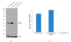
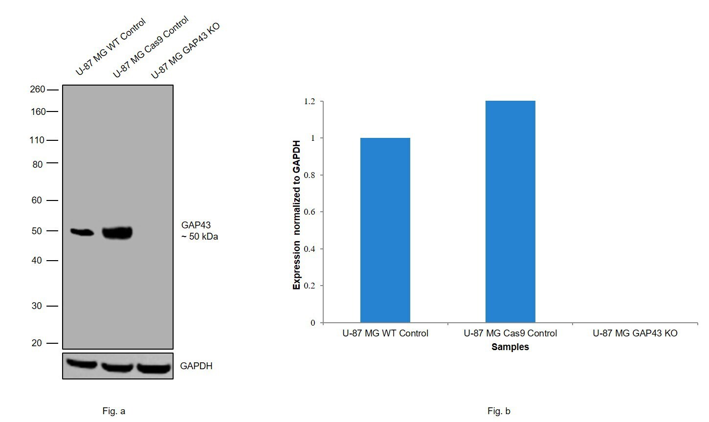
- Knockout of GAP43 was achieved by CRISPR-Cas9 genome editing. Western blot analysis of GAP43 was performed by loading 20 µg of U-87 MG wild type (Lane 1), U-87 MG CAS9 (Lane 2), U-87 MG GAP43 KO whole cell extracts. The blot was probed with Anti-GAP43 Polyclonal Antibody (Product # PA5-79299) using 1:2000 dilution and Goat anti-Rabbit IgG (H+L), Superclonal™ Recombinant Secondary Antibody, HRP (Product # A27036) using 1:4000 dilution. Loss of signal upon CRISPR mediated knockout (KO) confirms that antibody is specific to GAP43.
- Submitted by
- Invitrogen Antibodies (provider)
- Main image
- Experimental details
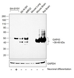
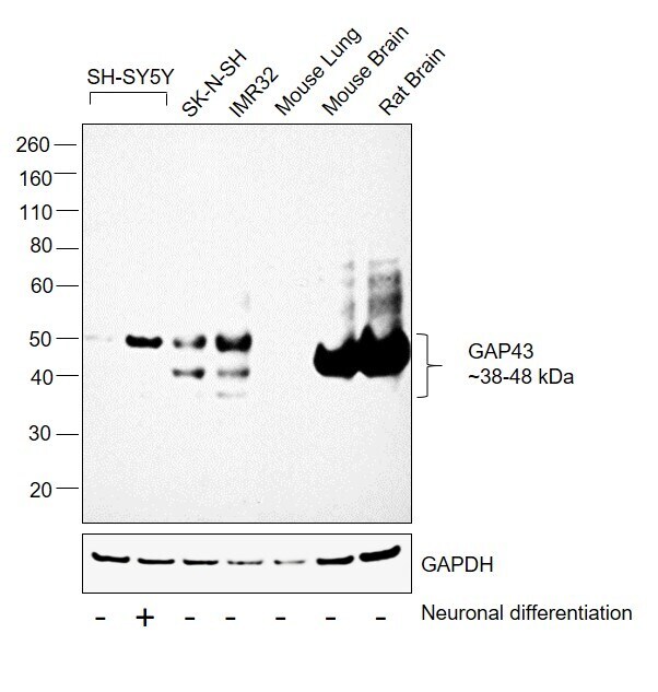
- Western blot was performed using Anti-GAP43 Polyclonal Antibody(Product # PA5-79299) and a 38-48kDa band corresponding to GAP43 was observed across cell lines and tissues tested except in Mouse Lung which is reported to be low to negative for GAP43 expression. An increase in GAP43 expression was observed in SH-SY5Y on neuronal differentiation. Membrane enriched extracts (30 µg lysate) of SH-SY5Y (Lane 1), SH-SY5Y differentiated to neurons (Lane 2), SK-N-SH (Lane 3), IMR-32 (Lane 4), Mouse Lung (Lane 5), Mouse Brain (Lane 6) and Rat Brain (Lane 7) were electrophoresed using NuPAGE™ 4-12% Bis-Tris Protein Gel (Product # NP0322BOX). Resolved proteins were then transferred onto a Nitrocellulose membrane (Product # IB23001) by iBlot® 2 Dry Blotting System (Product # IB21001). The blot was probed with the primary antibody (0.5ug/ml) and detected by chemiluminescence with Goat anti-Rabbit IgG (H+L) Superclonal™ Recombinant Secondary Antibody, HRP (Product # A27036,1:4000 dilution) using the iBright FL 1000 (Product # A32752). Chemiluminescent detection was performed using Novex® ECL Chemiluminescent Substrate Reagent Kit (Product # WP20005).
Supportive validation
- Submitted by
- Invitrogen Antibodies (provider)
- Main image
- Experimental details
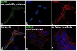
- Immunofluorescence analysis of GAP43 was performed using 70% confluent log phase SH-SY5Y cells differentiated to neurons and undifferentiated cells. The cells were fixed with 4% paraformaldehyde for 10 minutes, permeabilized with 0.1% Triton™ X-100 for 15 minutes, and blocked with 2% BSA for 45 minutes at room temperature. The cells were labeled with GAP43 Polyclonal Antibody (Product # PA5-79299) at 5 µg/mL in 0.1% BSA, incubated at 4-degree Celsius overnight and then labeled with Donkey anti-Rabbit IgG (H+L) Highly Cross-Adsorbed Secondary Antibody, Alexa Fluor Plus 488 (Product # A32790), (1:2000 dilution), for 45 minutes at room temperature (Panel a: Green). Nuclei (Panel b: Blue) were stained with ProLong™ Diamond Antifade Mountant with DAPI (Product # P36962). F-actin (Panel c: Red) was stained with Rhodamine Phalloidin (Product # R415, 1:300). Panel d represents the merged image showing Plasma membrane and cytoplasmic localization of the neuronal differentiated SH-SY5Y cells. Panel e represents undifferentiated SH-SY5Y cells with no expression for GAP43. Panel f represents control cells with no primary antibody to assess the background. The images were captured at 60X magnification.
Supportive validation
- Submitted by
- Invitrogen Antibodies (provider)
- Main image
- Experimental details
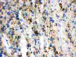
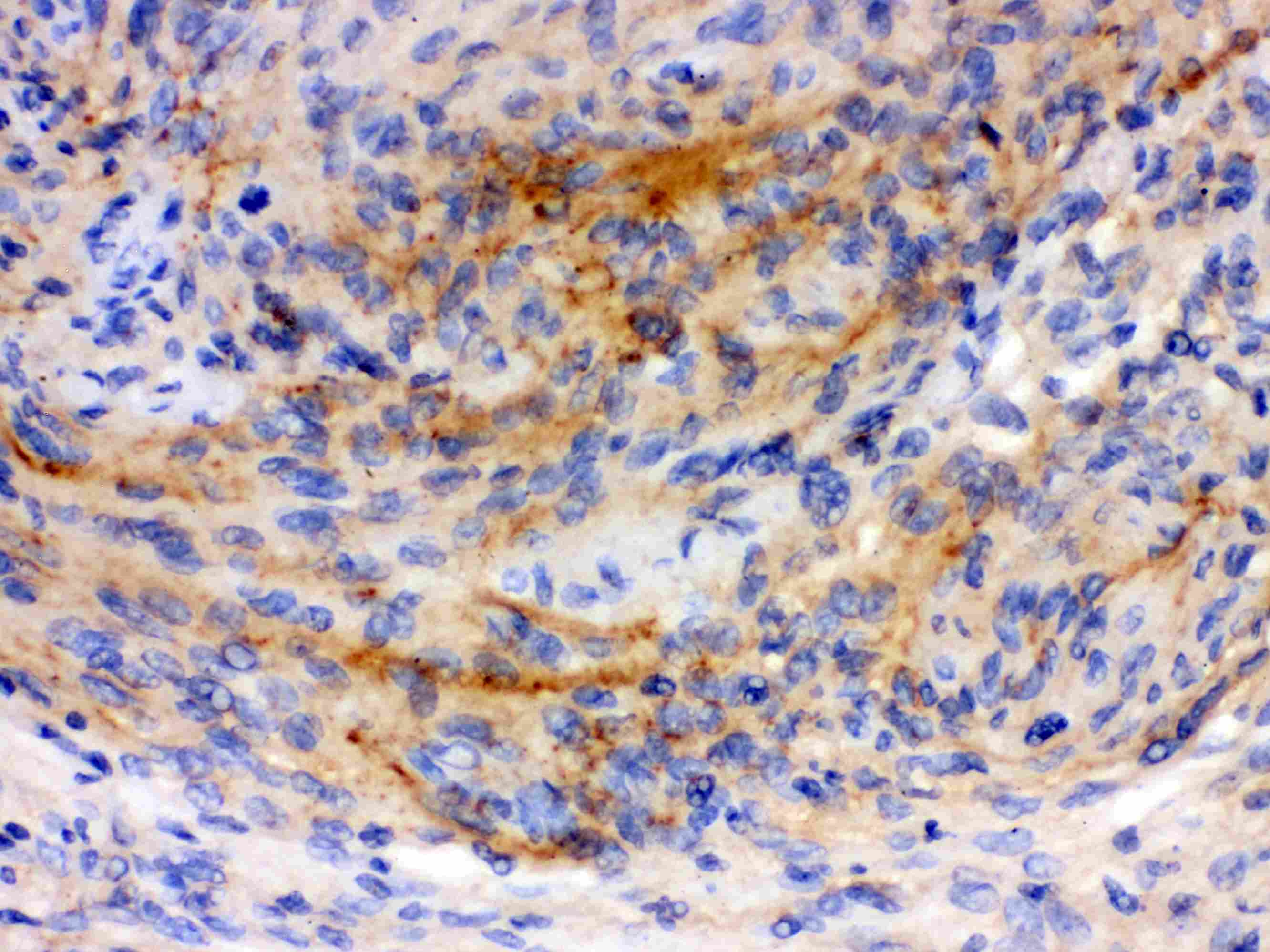
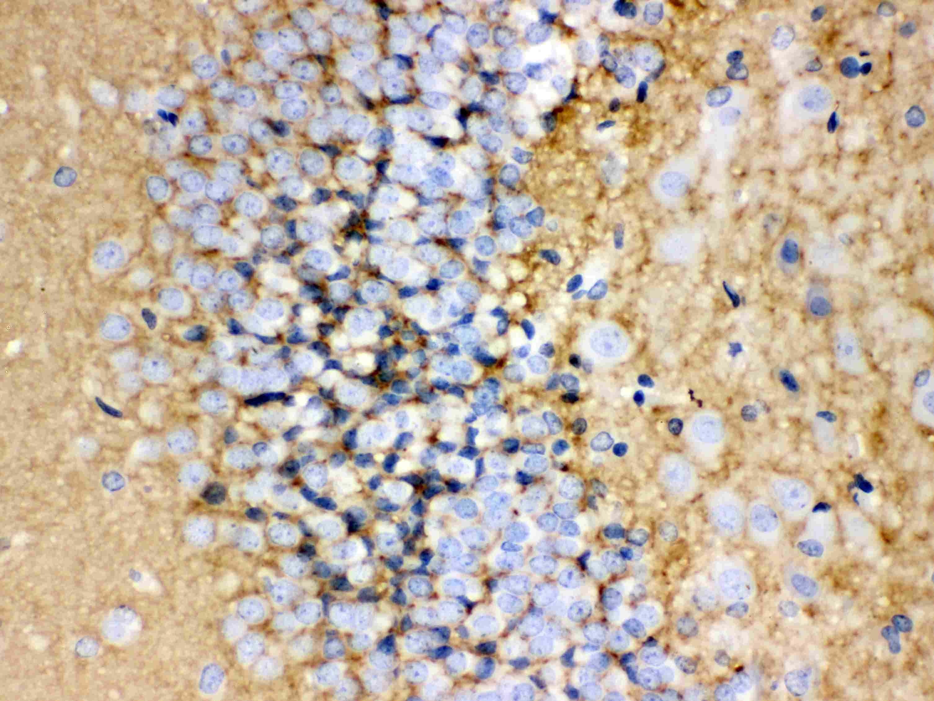
- Immunohistochemistry analysis of GAP43 on paraffin-embedded human glioma tissue. Sample was incubated with GAP43 polyclonal antibody (Product# PA5-79299).
- Submitted by
- Invitrogen Antibodies (provider)
- Main image
- Experimental details
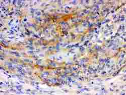
- Immunohistochemistry analysis of GAP43 on paraffin-embedded human meningioma tissue. Sample was incubated with GAP43 polyclonal antibody (Product# PA5-79299).
- Submitted by
- Invitrogen Antibodies (provider)
- Main image
- Experimental details
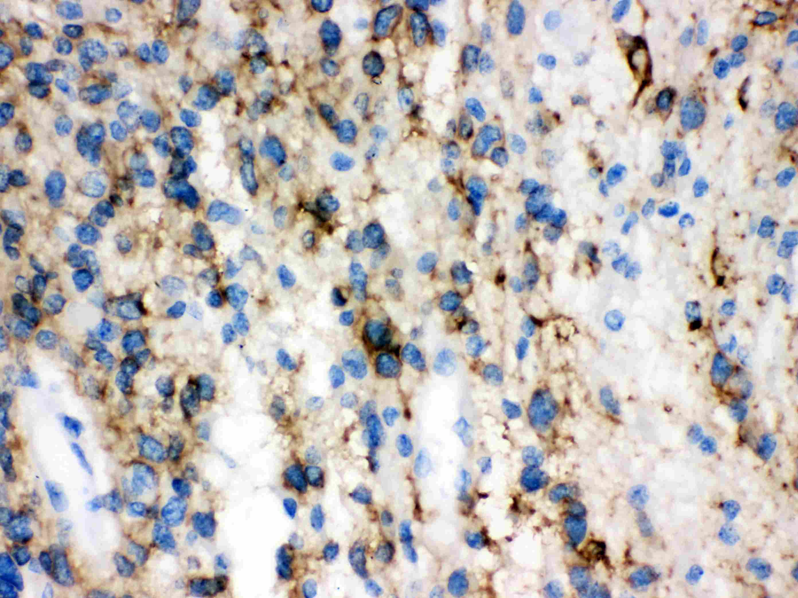
- Immunohistochemistry analysis of GAP43 on paraffin-embedded rat brain tissue. Sample was incubated with GAP43 polyclonal antibody (Product# PA5-79299).
Supportive validation
- Submitted by
- Invitrogen Antibodies (provider)
- Main image
- Experimental details
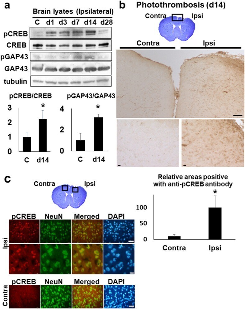
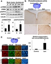
- Fig. 7 The activation of CREB and GAP in the stroke brain. a The western blot analyses with anti-CREB and -GAP43 antibody. The brain lysates (30 mug per each lane) of whole ipsi-lateral cortices at day 1, 3, 7, 14, and 28 were blotted with anti-CREB and anti-GAP43 antibody, respectively. C (control). The lower histogram shows the semi-quantification of band intensities of phosphorylated-CREB and -GAP43 relative to the total CREB and GAP43 protein (C vs. day 14), respectively (* p < 0.05, C vs. day14, Dunnett''s multiple comparison test). The value of control was made 1 for normalization. b The immunohistochemistry with ant-phosphorylated CREB (pCREB) antibody on the brain section, at day 14 post-stroke. The top picture shows the cresyl-violet stained bran section at day 14. Scale bar = 100 mum (upper panels), 10 mum (lower panels). The lower histogram shows the semi-quantification of DAB-stained areas positive with anti- pCREB antibody in ischemic brain sections (Contra- vs. Ipsi-lateral cortex) (* p < 0.05, Contra- vs. Ipsi-lateral, Student''s t-test). The value of ipsilateral brain was made 100 for normalization. Scale bar = 50 mum (upper panels), 10 mum (lower panels). c The immunofluorescence pictures of peri-ischemic regions on the Ipsi- and Contra-lateral cortex at day 14, with anti-phosphorylated CREB (pCREB) and anti-NeuN antibody, respectively. NeuN; Neuron-Specific Nuclear Protein. DAPI; 4'',6-diamidino-2-phenylindole. Scale bar = 50 mum (lower panels)