 Explore
 Validate
 Learn
Learn Western blot
Antibody data
- Antibody Data
- Antigen structure
- References [1]
- Comments [0]
- Validations
- Western blot [1]
- Immunohistochemistry [3]
Submit
Validation data
Reference
Comment
Report error
- Product number
- AF6259 - Provider product page

- Provider
- R&D Systems
- Product name
- Human/Mouse/Rat DARPP-32 Antibody
- Antibody type
- Polyclonal
- Description
- Immunogen affinity purified. Detects human, mouse, and rat DARPP-32 in Western blots.
- Reactivity
- Human, Mouse, Rat
- Host
- Goat
- Conjugate
- Unconjugated
- Antigen sequence
Q94D71- Isotype
- IgG
- Vial size
- 100 ug
- Concentration
- LYOPH
- Storage
- Use a manual defrost freezer and avoid repeated freeze-thaw cycles. 12 months from date of receipt, -20 to -70 °C as supplied. 1 month, 2 to 8 °C under sterile conditions after reconstitution. 6 months, -20 to -70 °C under sterile conditions after reconstitution.
Submitted references Early structural and functional plasticity alterations in a susceptibility period of DYT1 dystonia mouse striatum.
Maltese M, Stanic J, Tassone A, Sciamanna G, Ponterio G, Vanni V, Martella G, Imbriani P, Bonsi P, Mercuri NB, Gardoni F, Pisani A
eLife 2018 Mar 5;7
eLife 2018 Mar 5;7
No comments: Submit comment
Supportive validation
- Submitted by
- R&D Systems (provider)
- Main image
- Experimental details
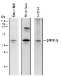
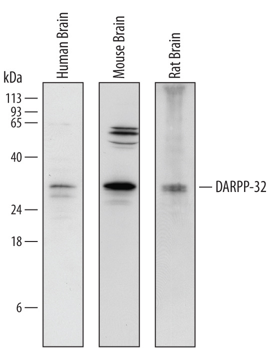
- Detection of Human, Mouse, and Rat DARPP-32 by Western Blot. Western blot shows lysates of human brain tissue, mouse brain tissue, and rat brain tissue. PVDF Membrane was probed with 1 µg/mL of Goat Anti-Human/Mouse/Rat DARPP-32 Antigen Affinity-purified Polyclonal Antibody (Catalog # AF6259) followed by HRP-conjugated Anti-Goat IgG Secondary Antibody (Catalog # HAF109). A specific band was detected for DARPP-32 at approximately 32 - 35 kDa (as indicated). This experiment was conducted under reducing conditions and using Immunoblot Buffer Group 1.
Supportive validation
- Submitted by
- R&D Systems (provider)
- Main image

- Experimental details
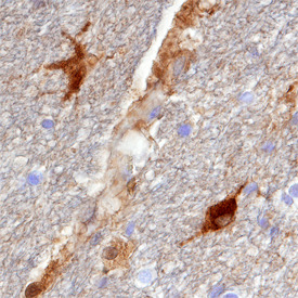
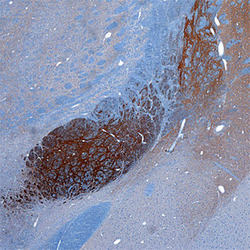
- DARPP-32 in Human Brain. DARPP-32 was detected in immersion fixed paraffin-embedded sections of human brain (hippocampus) using Goat Anti-Human/Mouse/Rat DARPP-32 Antigen Affinity-purified Polyclonal Antibody (Catalog # AF6259) at 15 µg/mL overnight at 4 °C. Tissue was stained using the Anti-Goat HRP-DAB Cell & Tissue Staining Kit (brown; Catalog # CTS008) and counterstained with hematoxylin (blue). Specific staining was localized to neurons and glial cells. View our protocol for Chromogenic IHC Staining of Paraffin-embedded Tissue Sections.
- Submitted by
- R&D Systems (provider)
- Main image
- Experimental details
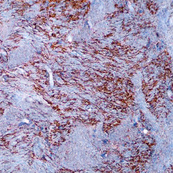
- DARPP-32 in Mouse Brain. DARPP-32 was detected in perfusion fixed frozen sections of mouse brain (caudate putamen) using Goat Anti-Human/Mouse/Rat DARPP-32 Antigen Affinity-purified Polyclonal Antibody (Catalog # AF6259) at 15 µg/mL overnight at 4 °C. Tissue was stained using the Anti-Goat HRP-DAB Cell & Tissue Staining Kit (brown; Catalog # CTS008) and counterstained with hematoxylin (blue). Specific staining was localized to neuronal processes. View our protocol for Chromogenic IHC Staining of Frozen Tissue Sections.

- Submitted by
- R&D Systems (provider)
- Main image
- Experimental details
- DARPP-32 in Rat Brain. DARPP-32 was detected in perfusion fixed frozen sections of rat brain (globus pallidus) using Goat Anti-Human/Mouse/Rat DARPP-32 Antigen Affinity-purified Polyclonal Antibody (Catalog # AF6259) at 15 µg/mL overnight at 4 °C. Tissue was stained using the Anti-Goat HRP-DAB Cell & Tissue Staining Kit (brown; Catalog # CTS008) and counterstained with hematoxylin (blue). Specific staining was localized to neuronal processes. View our protocol for Chromogenic IHC Staining of Frozen Tissue Sections.
