 Explore
 Validate
 Learn
Learn Western blot
Antibody data
- Antibody Data
- Antigen structure
- References [4]
- Comments [0]
- Validations
- Western blot [4]
- Immunohistochemistry [1]
Submit
Validation data
Reference
Comment
Report error
- Product number
- GTX102055 - Provider product page

- Provider
- GeneTex
- Proper citation
- GeneTex Cat#GTX102055, RRID:AB_10618726
- Product name
- MFN2 antibody [N1N2], N-term
- Antibody type
- Polyclonal
- Reactivity
- Human, Mouse
- Host
- Rabbit
Submitted references Laminar shear stress promotes mitochondrial homeostasis in endothelial cells.
Aqueous extracts of Paeonia suffruticosa modulates mitochondrial proteostasis by reactive oxygen species-induced endoplasmic reticulum stress in pancreatic cancer cells.
Enhancement of Mitochondrial Transfer by Antioxidants in Human Mesenchymal Stem Cells.
Metabolic Stress-Induced Phosphorylation of KAP1 Ser473 Blocks Mitochondrial Fusion in Breast Cancer Cells.
Wu LH, Chang HC, Ting PC, Wang DL
Journal of cellular physiology 2018 Jun;233(6):5058-5069
Journal of cellular physiology 2018 Jun;233(6):5058-5069
Aqueous extracts of Paeonia suffruticosa modulates mitochondrial proteostasis by reactive oxygen species-induced endoplasmic reticulum stress in pancreatic cancer cells.
Liu YH, Weng YP, Tsai HY, Chen CJ, Lee DY, Hsieh CL, Wu YC, Lin JY
Phytomedicine : international journal of phytotherapy and phytopharmacology 2018 Jul 15;46:184-192
Phytomedicine : international journal of phytotherapy and phytopharmacology 2018 Jul 15;46:184-192
Enhancement of Mitochondrial Transfer by Antioxidants in Human Mesenchymal Stem Cells.
Li CJ, Chen PK, Sun LY, Pang CY
Oxidative medicine and cellular longevity 2017;2017:8510805
Oxidative medicine and cellular longevity 2017;2017:8510805
Metabolic Stress-Induced Phosphorylation of KAP1 Ser473 Blocks Mitochondrial Fusion in Breast Cancer Cells.
Cheng CT, Kuo CY, Ouyang C, Li CF, Chung Y, Chan DC, Kung HJ, Ann DK
Cancer research 2016 Sep 1;76(17):5006-18
Cancer research 2016 Sep 1;76(17):5006-18
No comments: Submit comment
Supportive validation
- Submitted by
- GeneTex (provider)
- Main image

- Experimental details
- Sample (30 ug of whole cell lysate) A: A549 7.5% SDS PAGE GTX102055 diluted at 1:1000
- Validation comment
- WB

- Submitted by
- GeneTex (provider)
- Main image
- Experimental details
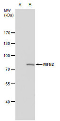
- MFN2 antibody [N1N2], N-term detects MFN2 protein by western blot analysis.A. 30 ?g 293T whole cell extractB. 30 ?g whole cell extract of human MFN2 transfected 293T cells7.5 % SDS-PAGEMFN2 antibody [N1N2], N-term (GTX102055) dilution: 1:5000
- Validation comment
- WB

- Submitted by
- GeneTex (provider)
- Main image

- Experimental details
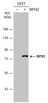
- MFN2 antibody [N1N2], N-term detects MFN2 protein by western blot analysis.A. 30 ?g 293T whole cell extractB. 30 ?g whole cell extract of human MFN2 transfected 293T cells7.5% SDS-PAGEMFN2 antibody [N1N2], N-term (GTX102055) dilution: 1:5000 The HRP-conjugated anti-rabbit IgG antibody (GTX213110-01) was used to detect the primary antibody.

- Submitted by
- GeneTex (provider)
- Main image

- Experimental details
- Non-transfected (¡V) and transfected (+) 293T whole cell extracts (30 ?g) were separated by 7.5% SDS-PAGE, and the membrane was blotted with MFN2 antibody [N1N2], N-term (GTX102055) diluted at 1:1000. The HRP-conjugated anti-rabbit IgG antibody (GTX213110-01) was used to detect the primary antibody.
Supportive validation
- Submitted by
- GeneTex (provider)
- Main image
- Experimental details
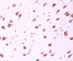
- MFN2 antibody [N1N2], N-term detects MFN2 protein at cytosol on mouse fore brain by immunohistochemical analysis. Sample: Paraffin-embedded mouse fore brain. MFN2 antibody [N1N2], N-term (GTX102055) dilution: 1:500.
