 Explore
 Validate
 Learn
Learn Western blot
 Immunoprecipitation
Antibody data
- Antibody Data
- Antigen structure
- References [0]
- Comments [0]
- Validations
- Western blot [3]
- Immunocytochemistry [1]
- Immunohistochemistry [1]
- Other assay [1]
Submit
Validation data
Reference
Comment
Report error
- Product number
- PA5-118059 - Provider product page

- Provider
- Invitrogen Antibodies
- Product name
- MFN2 Polyclonal Antibody
- Antibody type
- Polyclonal
- Antigen
- Other
- Reactivity
- Human, Mouse, Rat
- Host
- Rabbit
- Isotype
- IgG
- Vial size
- 100 µL
- Storage
- -20° C, Avoid Freeze/Thaw Cycles
No comments: Submit comment
Supportive validation
- Submitted by
- Invitrogen Antibodies (provider)
- Main image
- Experimental details
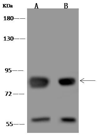
- Western Blot using MFN2 Polyclonal Antibody (Product # PA5-118059) at 1:500 dilution. Lane A: Jurkat Whole Cell Lysate. Lysates/proteins at 30 μg per lane. Secondary antibody: Goat Anti-Rabbit IgG (H+L)/HRP at 1:10,000 dilution. Developed using the ECL technique. Performed under reducing conditions. Predicted band size: 86 kDa. Observed band size: 86 kDa. (We are unsure of the identity of these extra bands).
- Submitted by
- Invitrogen Antibodies (provider)
- Main image
- Experimental details
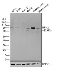
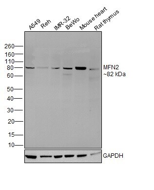
- Western blot was performed using Anti-MFN2 Polyclonal Antibody (Product # PA5-118059) and a 82 kDa band corresponding to MFN2 was observed across cell lines and tissues tested. Whole cell extracts (30 µg lysate) of A549 (Lane 1), Reh (Lane 2), IMR-32 (Lane 3), BeWo (Lane 4); and tissue extracts of Mouse Heart (Lane 5) and Rat Thymus (Lane 6) were electrophoresed using NuPAGE™ 10% Bis-Tris Protein Gel (Product # NP0301BOX). Resolved proteins were then transferred onto a nitrocellulose membrane (Product # IB23001) by iBlot® 2 Dry Blotting System (Product # IB21001). The blot was probed with the primary antibody (1:1000 dilution) and detected by chemiluminescence with Goat anti-Rabbit IgG (H+L) Superclonal™ Recombinant Secondary Antibody, HRP (Product # A27036,1:20000 dilution) using the iBright™ FL1500 Imaging System (Product # A44115). Chemiluminescent detection was performed using SuperSignal™ West Atto Ultimate Sensitivity Substrate (Product # A38556).
- Submitted by
- Invitrogen Antibodies (provider)
- Main image
- Experimental details
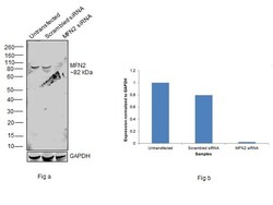
- Knockdown of MFN2 was achieved by transfecting A549 with MFN2 specific siRNAs (Silencer® select Product # S502615, S19262). Western blot analysis (Fig. a) was performed using Whole cell extracts from the MFN2 knockdown cells (lane 3), non-targeting scrambled siRNA transfected cells (lane 2) and untransfected cells (lane 1). The blot was probed with MFN2 Polyclonal Antibody (Product # PA5-118059, 1:1000 dilution) and Goat anti-Rabbit IgG (H+L) Superclonal™ Recombinant Secondary Antibody, HRP (Product # A27036, 1:20000 dilution). Densitometric analysis of this western blot is shown in histogram (Fig. b). Decrease in signal upon siRNA mediated knock down confirms that antibody is specific to MFN2.

Supportive validation
- Submitted by
- Invitrogen Antibodies (provider)
- Main image
- Experimental details
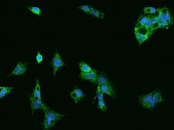
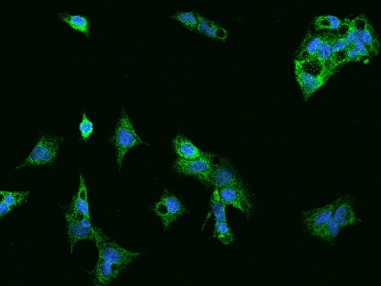
- Immunofluorescence staining of MFN2 in HepG2 cells. Cells were fixed with 4% PFA, permeabilzed with 0.1% Triton X-100 in PBS, blocked with 10% serum, and incubated with MFN2 Polyclonal Antibody (Product # PA5-118059, 1:200) at 4°C overnight. Then cells were stained with the Alexa Fluor®488-conjugated Goat Anti-rabbit IgG secondary antibody (green) and counterstained with DAPI (blue). Positive staining was localized to cytoplasm.
Supportive validation
- Submitted by
- Invitrogen Antibodies (provider)
- Main image
- Experimental details
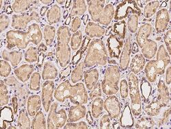
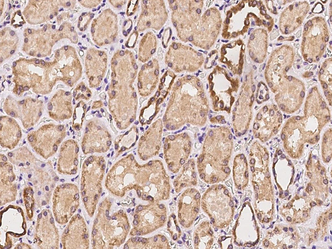
- Immunohistochemical staining of human MFN2 in human kidney with MFN2 Polyclonal Antibody (Product # PA5-118059, 1:100 dilution, formalin-fixed paraffin embedded sections).
Supportive validation
- Submitted by
- Invitrogen Antibodies (provider)
- Main image
- Experimental details
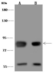
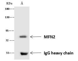
- MFN2 Immunoprecipitation using: Lane A: 0.5 mg HeLa Jurkat Whole Cell Lysate 4 µL with MFN2 Polyclonal Antibody (Product # PA5-118059) and 60 μg of Immunomagnetic beads Protein A/G. Primary antibody: MFN2 Polyclonal Antibody, at 1:100 dilution. Secondary antibody: Goat Anti-Rabbit IgG (H+L) /HRP at 1:10,000 dilution. Developed using the ECL technique. Performed under reducing conditions. Predicted band size: 86 kDa. Observed band size: 86 kDa.
