 Explore
 Validate
 Learn
LearnPA5-116092
antibody from Invitrogen Antibodies
Targeting: TUT1
FLJ21850, FLJ22267, FLJ22347, PAPD2, RBM21, TENT1, TUTase
 Western blot
Antibody data
- Antibody Data
- Antigen structure
- References [0]
- Comments [0]
- Validations
- Western blot [1]
- Immunocytochemistry [1]
- Immunohistochemistry [4]
Submit
Validation data
Reference
Comment
Report error
- Product number
- PA5-116092 - Provider product page

- Provider
- Invitrogen Antibodies
- Product name
- TUT1 Polyclonal Antibody
- Antibody type
- Polyclonal
- Antigen
- Synthetic peptide
- Reactivity
- Human, Mouse, Rat
- Host
- Rabbit
- Isotype
- IgG
- Vial size
- 100 µL
- Concentration
- 1 mg/mL
- Storage
- -20°C
No comments: Submit comment
Supportive validation
- Submitted by
- Invitrogen Antibodies (provider)
- Main image
- Experimental details
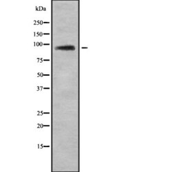
- Western blot analysis of TUT1 in 3T3 whole cells lysates. Samples were incubated with polyclonal antibody (Product # PA5-116092).

Supportive validation
- Submitted by
- Invitrogen Antibodies (provider)
- Main image

- Experimental details
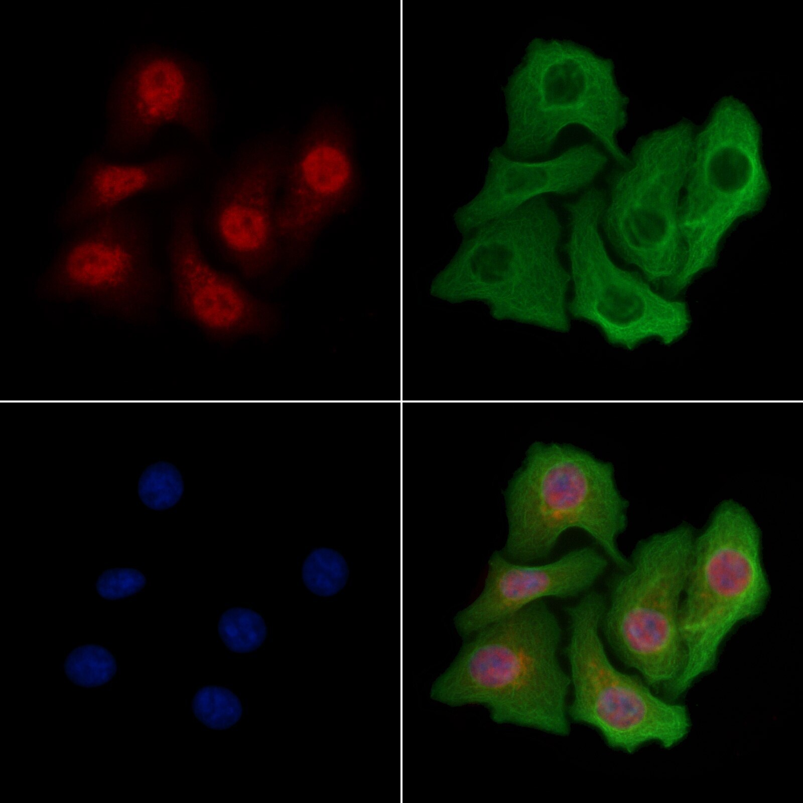
- Immunocytochemistry analysis of TUT1 in HeLa cells. Samples were treated with PFA, permeabilized in 0.1% Triton X-100, blocked in 10% serum (45 min at 25°C), and incubated with polyclonal antibody (Product # PA5-116092). Secondary staining was applied with mouse anti-beta tubulin (1 hr at 37°, AlexaFluor 594 conjugated goat anti-rabbit IgG (Red), AlexaFluor 488 conjugated goat anti-mouse IgG (Green) and DAPI.
Supportive validation
- Submitted by
- Invitrogen Antibodies (provider)
- Main image
- Experimental details
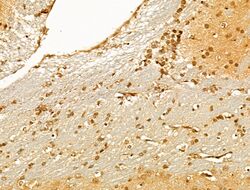
- Immunohistochemistry analysis of TUT1 in mouse brain tissue. Samples were treated with formaldehyde and treated with citrate buffer for antigen retrieval, blocked, and incubated (1.5 hours at 22°C) with polyclonal antibody (Product # PA5-116092) at a dilution of 1:100. Secondary staining was applied with HRP conjugated anti-Rabbit.

- Submitted by
- Invitrogen Antibodies (provider)
- Main image
- Experimental details
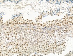
- Immunohistochemistry analysis of TUT1 in mouse testis tissue. Samples were treated with formaldehyde and treated with citrate buffer for antigen retrieval, blocked, and incubated (1.5 hours at 22°C) with polyclonal antibody (Product # PA5-116092) at a dilution of 1:100. Secondary staining was applied with HRP conjugated anti-Rabbit.

- Submitted by
- Invitrogen Antibodies (provider)
- Main image

- Experimental details
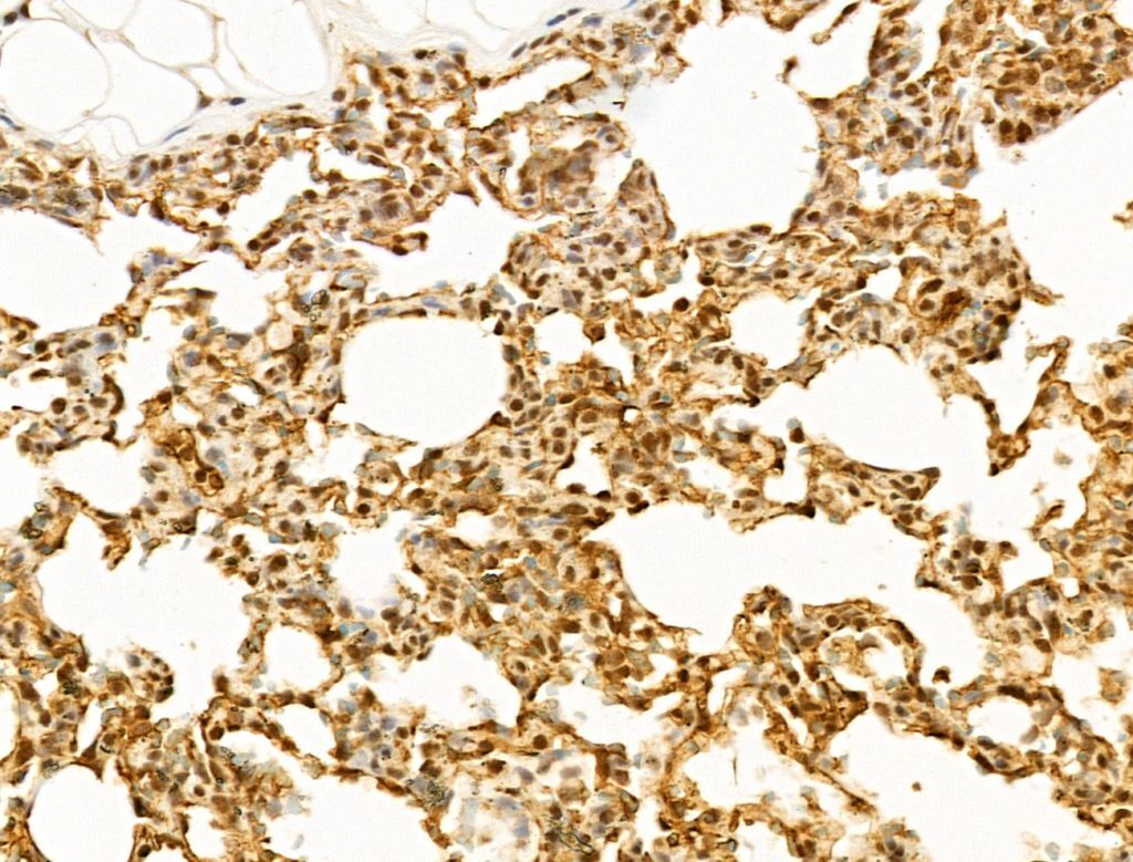
- Immunohistochemistry analysis of TUT1 in rat lung tissue. Samples were treated with formaldehyde and treated with citrate buffer for antigen retrieval, blocked, and incubated (1.5 hours at 22°C) with polyclonal antibody (Product # PA5-116092) at a dilution of 1:100. Secondary staining was applied with HRP conjugated anti-Rabbit.
- Submitted by
- Invitrogen Antibodies (provider)
- Main image
- Experimental details
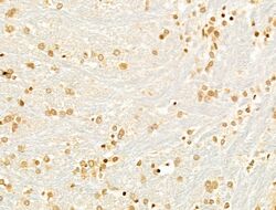
- Immunohistochemistry analysis of TUT1 in rat brain tissue. Samples were treated with formaldehyde and treated with citrate buffer for antigen retrieval, blocked, and incubated (1.5 hours at 22°C) with polyclonal antibody (Product # PA5-116092) at a dilution of 1:100. Secondary staining was applied with HRP conjugated anti-Rabbit.
