Antibody data
- Antibody Data
- Antigen structure
- References [0]
- Comments [0]
- Validations
- Western blot [1]
- Immunohistochemistry [1]
- Other assay [1]
Submit
Validation data
Reference
Comment
Report error
- Product number
- PA5-54271 - Provider product page

- Provider
- Invitrogen Antibodies
- Product name
- NEK1 Polyclonal Antibody
- Antibody type
- Polyclonal
- Antigen
- Recombinant full-length protein
- Description
- Immunogen sequence: NLKAQEDEKG KQNLSDTFEI NVHEDAKEHE KEKSVSSDRK KWEAGGQLVI PLDELTLDTS FSTTERHTVG EVIKLGPNGS PRRAWGKSPT DSVLKILGEA ELQLQTELLE NTTIRSEISP EGEKYKPLIT GEKKVQCISH EINPSA Highest antigen sequence identity to the following orthologs: Mouse - 72%, Rat - 71%.
- Reactivity
- Human
- Host
- Rabbit
- Isotype
- IgG
- Vial size
- 100 µL
- Concentration
- 0.7 mg/mL
- Storage
- Store at 4°C short term. For long term storage, store at -20°C, avoiding freeze/thaw cycles.
No comments: Submit comment
Supportive validation
- Submitted by
- Invitrogen Antibodies (provider)
- Main image

- Experimental details
- Western blot of Nek1 was performed by loading 100 µg of WT (lane 1) and NEK1 CRISPR KO (lane 2) HeLa cell lysates in RIPA buffer onto a 5-16% gradient polyacrylamide gel. Proteins on the blots were visualized with Ponceau staining (below immunoblot). Proteins were transferred to nitrocellulose membrane and blocked in 5% milk for 1 hr. Nek1 was detected at approximately 143 kDa (designated by the black arrow) using a Nek1 polyclonal antibody (Product # PA5-54271) at a dilution of 1:1000 in 5% BSA in TBS with 0.1% Tween 20 (TBST) overnight at 4 deg. The peroxidase-conjugated secondary antibody (Product # 65-6120) was diluted to 0.2 µg/mL in TBST with 5% milk for 1 hr. Chemiluminescent detection was performed using Pierce ECL Western Blotting Substrate (Product # 32106). Data courtesy of YCharOS Inc., an open science company with the mission of characterizing commercially available antibodies using knockout validation.
Supportive validation
- Submitted by
- Invitrogen Antibodies (provider)
- Main image

- Experimental details
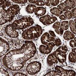
- Immunohistochemical staining of NEK1 in human stomach tissue shows strong cytoplasmic positivity in glandular cells. Samples were probed using a NEK1 Polyclonal Antibody (Product # PA5-54271).
Supportive validation
- Submitted by
- Invitrogen Antibodies (provider)
- Main image

- Experimental details
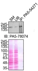
- Immunoprecipitation of NEK1 was performed on HeLa cell lysates. Antibody-bead conjugates were prepared by adding 1 µg of NEK1 polyclonal antibody (Product # PA5-54271) with 30 µL of protein A -Sepharose beads and rocked overnight at 4 deg. 1 µg of lysate was incubated with antibody-bead conjugate for 2 hrs at 4 deg. After multiple washes, 10% starting material (SM), 10% unbound fraction (UB) and immunoprecipitated fraction (IP) were processed for immunoblot using a different NEK1 polyclonal antibody (Product # PA5-78074). Ponceau stained transfer of blot is shown. Data courtesy of YCharOS Inc., an open science company with the mission of characterizing commercially available antibodies using knockout validation.
 Explore
 Validate
 Learn
Learn Western blot
 Immunoprecipitation