Explore
 Validate
 Learn
Learn Western blot
Antibody data
- Antibody Data
- Antigen structure
- References [1]
- Comments [0]
- Validations
- Western blot [2]
- Immunohistochemistry [12]
- Other assay [1]
Submit
Validation data
Reference
Comment
Report error
- Product number
- MA5-24650 - Provider product page

- Provider
- Invitrogen Antibodies
- Product name
- Laminin alpha-4 Monoclonal Antibody (CL3183)
- Antibody type
- Monoclonal
- Antigen
- Recombinant full-length protein
- Description
- Immunogen sequence: ENLLNQAREL QAKAESSSDE AVADTSRRVG GALARKSALK TRLSDAVKQL QAAERGDAQQ RLGQSRLITE EANRTTMEVQ QATAPMANNL TNWSQNLQHF DSSAYNTAVN SARDAVRNLT EVVPQLLDQL RTVEQKRPAS
- Antibody clone number
- CL3183
- Concentration
- 1 mg/mL
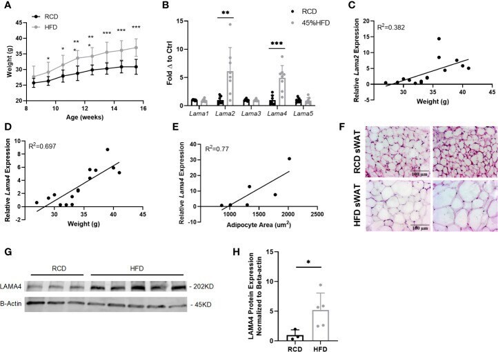
Submitted references Laminin-α4 Is Upregulated in Both Human and Murine Models of Obesity.
Goddi A, Carmona A, Schroedl L, White JM, Piron MJ, De Leon A, Casimiro I, Hoffman A, Gonzalez Porras MA, Brey EM, Brady MJ, Cohen RN
Frontiers in endocrinology 2021;12:698621
Frontiers in endocrinology 2021;12:698621
No comments: Submit comment
Supportive validation
- Submitted by
- Invitrogen Antibodies (provider)
- Main image

- Experimental details
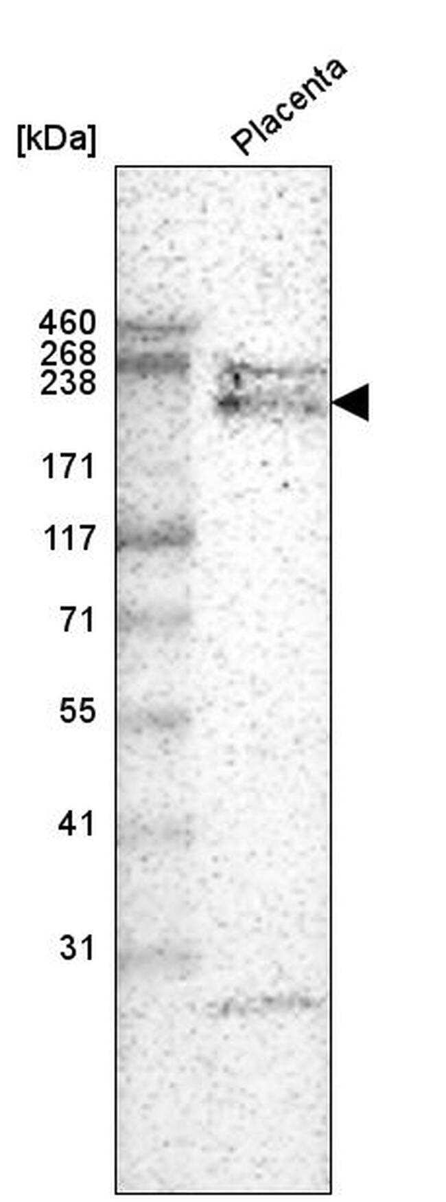
- Western blot analysis of Laminin alpha-4 in human placenta tissue using a Laminin alpha-4 Monoclonal Antibody (CL3183) (Product # MA5-24650).
- Submitted by
- Invitrogen Antibodies (provider)
- Main image

- Experimental details
- Western blot analysis of purified human recombinant Laminin-411, Laminin-421, Laminin-511, Laminin-121, Laminin-221 and Laminin-332 using a Laminin alpha-4 Monoclonal Antibody (CL3183) (Product # MA5-24650).

Supportive validation
- Submitted by
- Invitrogen Antibodies (provider)
- Main image
- Experimental details
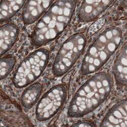
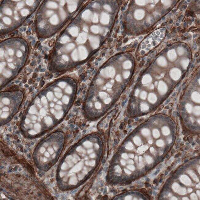
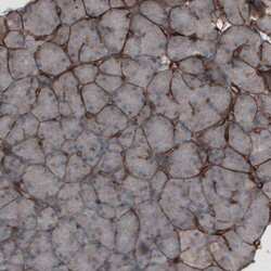
- Immunohistochemical staining of Laminin alpha-4 in human colon tissue shows strong immunoreactivity in basement membrane of glandular epithelium. Samples were probed using a Laminin alpha-4 Monoclonal Antibody (Product # MA5-24650).
- Submitted by
- Invitrogen Antibodies (provider)
- Main image
- Experimental details
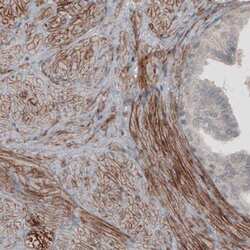
- Immunohistochemical staining of Laminin alpha-4 in human prostate shows moderate membranous positivity in smooth muscle cells. Samples were probed using a Laminin alpha-4 Monoclonal Antibody (Product # MA5-24650).

- Submitted by
- Invitrogen Antibodies (provider)
- Main image
- Experimental details
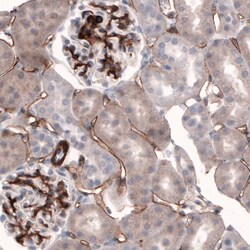
- Immunohistochemical staining of Laminin alpha-4 in human pancreatic tissue shows strong immunoreactivity in basement membrane of glandular epithelium. Samples were probed using a Laminin alpha-4 Monoclonal Antibody (Product # MA5-24650).

- Submitted by
- Invitrogen Antibodies (provider)
- Main image
- Experimental details
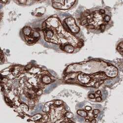
- Immunohistochemical staining of Laminin alpha-4 in human placenta shows strong positivity in basement membrane and endothelium. Samples were probed using a Laminin alpha-4 Monoclonal Antibody (Product # MA5-24650).

- Submitted by
- Invitrogen Antibodies (provider)
- Main image

- Experimental details
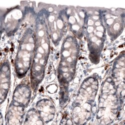
- Immunohistochemical staining of Laminin alpha-4 in human lymph node tissue shows absence of immunoreactivity in lymphoid tissue (negative control). Samples were probed using a Laminin alpha-4 Monoclonal Antibody (Product # MA5-24650).

- Submitted by
- Invitrogen Antibodies (provider)
- Main image
- Experimental details
- Immunohistochemical staining of Laminin alpha-4 in rat rectal tissue shows immunoreactivity in the basement membrane of glandular epithelium. Samples were probed using a Laminin alpha-4 Monoclonal Antibody (Product # MA5-24650).

- Submitted by
- Invitrogen Antibodies (provider)
- Main image
- Experimental details
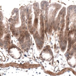
- Immunohistochemical staining of Laminin alpha-4 in mouse stomach tissue shows positivity in the basement membrane of glandular epithelium. Samples were probed using a Laminin alpha-4 Monoclonal Antibody (Product # MA5-24650).

- Submitted by
- Invitrogen Antibodies (provider)
- Main image
- Experimental details
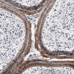
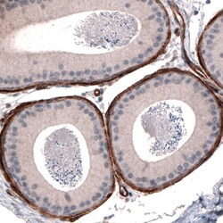
- Immunohistochemical staining of Laminin alpha-4 in mouse epididymis shows immunoreactivity in smooth muscle in the lamina propria. Samples were probed using a Laminin alpha-4 Monoclonal Antibody (Product # MA5-24650).

- Submitted by
- Invitrogen Antibodies (provider)
- Main image
- Experimental details
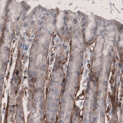
- Immunohistochemical staining of Laminin alpha-4 in rat stomach tissue shows immunoreactivity in the basement membrane of glandular epithelium. Samples were probed using a Laminin alpha-4 Monoclonal Antibody (Product # MA5-24650).

- Submitted by
- Invitrogen Antibodies (provider)
- Main image
- Experimental details
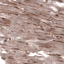
- Immunohistochemical staining of Laminin alpha-4 in rat heart muscle shows membranous positivity in cardiomyocytes. Samples were probed using a Laminin alpha-4 Monoclonal Antibody (Product # MA5-24650).

- Submitted by
- Invitrogen Antibodies (provider)
- Main image
- Experimental details
- Immunohistochemical staining of Laminin alpha-4 in rat kidney tissue shows basement membrane immunoreactivity in renal tubules and glomeruli. Samples were probed using a Laminin alpha-4 Monoclonal Antibody (Product # MA5-24650).

- Submitted by
- Invitrogen Antibodies (provider)
- Main image
- Experimental details
- Immunohistochemical staining of Laminin alpha-4 in rat epididymis shows strong immunoreactivity in lamina propria. Samples were probed using a Laminin alpha-4 Monoclonal Antibody (Product # MA5-24650).

Supportive validation
- Submitted by
- Invitrogen Antibodies (provider)
- Main image

- Experimental details
- Figure 1 Gene expression of laminin-alpha chains in sWAT of HFD fed mice. (A) Average weekly weights of mice on regular chow diet (RCD) and 45% High Fat Diet (HFD) for 8 weeks. RCD (n=7), HFD (n=8). (*,**,***) indicates p < 0.05, 0.01, 0.001, respectively. Data are Means +- SD. (B) Laminin-alpha chain mRNA expression in sWAT of mice after 8 weeks of dietary study normalized to the average of RCD control group. RCD (n=7), HFD (n=8). (C) Relative Lama2 mRNA expression normalized to RCD group graphed against weight (grams) for each mouse in 8-week dietary study (n=15). Simple linear regression analysis. (D) Relative Lama4 mRNA expression graphed against weight (grams) for each mouse in 8-week dietary study (n=15). Simple linear regression analysis. (E) Relative Lama4 mRNA expression graphed against adipocyte area (um 2 ) for one cohort of HFD and RCD mice (n=6). Simple linear regression analysis. (F) Representative images of H&E stained sWAT tissue sections of HFD and RCD mice after 8 weeks of dietary study. All images are 40X magnification. (G) Protein expression of LAMA4 and loading control B-actin in sWAT of mice placed on RCD or 45% HFD for 8 weeks was assessed by western blot. Original blot images can be found in the supplementary file. (H) LAMA4 signal from the western blot was quantified and normalized to the loading control signal. Values are shown as fold changes in comparison to the average of the RCD group. RCD (n=3), HFD (n=5).