Explore
 Validate
 Learn
Learn Western blot
 ELISA
ELISAAntibody data
- Antibody Data
- Antigen structure
- References [0]
- Comments [0]
- Validations
- Western blot [1]
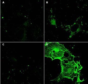
- Immunocytochemistry [1]
Submit
Validation data
Reference
Comment
Report error
- Product number
- AP09133PU-N - Provider product page

- Provider
- Acris Antibodies GmbH
- Proper citation
- Acris Antibodies GmbH Cat#AP09133PU-N, RRID:AB_2035338
- Product name
- anti CD97 (1-512)
- Antibody type
- Polyclonal
- Antigen
- Recombinant protein corresponding to amino acids 1-512 (extracellular domain) of Mouse CD97 protein.
- Reactivity
- Mouse
- Host
- Rabbit
- Isotype
- IgG
- Vial size
- 0.5 mg
- Concentration
- 5.0 mg/ml (UV absorbance at 280 nm)
No comments: Submit comment
Supportive validation
- Submitted by
- Acris Antibodies GmbH (provider)
- Main image

- Experimental details
- Western blot using Protein A purified anti-CD97 antibody shows detection of bands corresponding to free Fc-CD97- (5EGF) (lower arrowhead) and Fc-CD97- (5EGF) present as a complex (upper arrowhead) in lysates from COS cells. The left lane contains lysate from cells transfected with control DNA. The right lane contains lysate from COS cells expressing Fc-CD97- (5EGF). No staining was noted from bone marrow lysates taken from CD97 knockout mice. The identity of the band at ~65 kDa appearing in all lanes is not known. The formation of the CD97 complex is currently under investigation. Approximately 10 µl of lysate was used in each lane. A 1:1,000 dilution of the primary antibody was used. The image was processed using a 10-sec exposure.

Supportive validation
- Submitted by
- Acris Antibodies GmbH (provider)
- Main image

- Experimental details
- Immunofluorescence Microscopy using Protein A purified anti-CD97 antibody shows staining of Fc-CD97-(5EGF) (panel D) in transfected COS cells. Panel A and C shows similar staining using pre-immune serum. Panel A and B show staining of mock transfected COS cells (no vector). A 1:1,500 dilution of the primary antibody was used.