 Explore
 Validate
 Learn
Learn Western blot
 Immunocytochemistry
Antibody data
- Antibody Data
- Antigen structure
- References [2]
- Comments [0]
- Validations
- Western blot [1]
- Immunohistochemistry [1]
Submit
Validation data
Reference
Comment
Report error
- Product number
- NBP1-86176 - Provider product page

- Provider
- Novus Biologicals
- Proper citation
- Novus Cat#NBP1-86176, RRID:AB_11037096
- Product name
- Rabbit Polyclonal SYTL4 Antibody
- Antibody type
- Polyclonal
- Description
- Immunogen affinity purified. Specificity of human SYTL4 antibody verified on a Protein Array containing target protein plus 383 other non-specific proteins.
- Reactivity
- Human
- Host
- Rabbit
- Isotype
- IgG
- Vial size
- 0.1 ml
- Storage
- Store at 4C short term. Aliquot and store at -20C long term. Avoid freeze-thaw cycles.
Submitted references Neurexin-1α contributes to insulin-containing secretory granule docking.
Hermansky-Pudlak syndrome protein complexes associate with phosphatidylinositol 4-kinase type II alpha in neuronal and non-neuronal cells.
Mosedale M, Egodage S, Calma RC, Chi NW, Chessler SD
The Journal of biological chemistry 2012 Feb 24;287(9):6350-61
The Journal of biological chemistry 2012 Feb 24;287(9):6350-61
Hermansky-Pudlak syndrome protein complexes associate with phosphatidylinositol 4-kinase type II alpha in neuronal and non-neuronal cells.
Salazar G, Zlatic S, Craige B, Peden AA, Pohl J, Faundez V
The Journal of biological chemistry 2009 Jan 16;284(3):1790-802
The Journal of biological chemistry 2009 Jan 16;284(3):1790-802
No comments: Submit comment
Supportive validation
- Submitted by
- Novus Biologicals (provider)
- Main image
- Experimental details
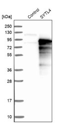
- Western Blot: SYTL4 Antibody [NBP1-86176] - Analysis in control (vector only transfected HEK293T lysate) and SYTL4 over-expression lysate (Co-expressed with a C-terminal myc-DDK tag (3.1 kDa) in mammalian HEK293T cells).

Supportive validation
- Submitted by
- Novus Biologicals (provider)
- Main image

- Experimental details
- Immunohistochemistry-Paraffin: SYTL4 Antibody [NBP1-86176] - Staining of human pancreas shows strong cytoplasmic positivity in exocrine glandular cells.
