 Explore
 Validate
 Learn
Learn Western blot
 Immunohistochemistry
Antibody data
- Antibody Data
- Antigen structure
- References [1]
- Comments [0]
- Validations
- Western blot [2]
- Immunocytochemistry [1]
- Immunohistochemistry [6]
Submit
Validation data
Reference
Comment
Report error
- Product number
- HPA015593 - Provider product page

- Provider
- Atlas Antibodies
- Proper citation
- Atlas Antibodies Cat#HPA015593, RRID:AB_2072217
- Product name
- Anti-NSRP1
- Antibody type
- Polyclonal
- Reactivity
- Human
- Host
- Rabbit
- Conjugate
- Unconjugated
- Antigen sequence
KREVGVQSSERNQDRKESSPNSRAKDKFLDQERSN
KMRNMAKDKERNQEKPSNSESSLGAKHRLTEEGQE
KGKEQERPPEAVSKFAKRNNEETVM- Isotype
- IgG
- Vial size
- 100 µl
- Storage
- Store at +4°C for short term storage. Long time storage is recommended at -20°C.
Submitted references Immunofluorescence and fluorescent-protein tagging show high correlation for protein localization in mammalian cells
Stadler C, Rexhepaj E, Singan V, Murphy R, Pepperkok R, Uhlén M, Simpson J, Lundberg E
Nature Methods 2013 February;10(4):315-323
Nature Methods 2013 February;10(4):315-323
No comments: Submit comment
Supportive validation
- Submitted by
- Atlas Antibodies (provider)
- Enhanced method
- Genetic validation
- Main image

- Experimental details
- Western blot analysis in HEK293 cells transfected with control siRNA, target specific siRNA probe #1 and #2, using Anti-NSRP1 antibody. Remaining relative intensity is presented. Loading control: Anti-GAPDH.

- Submitted by
- Atlas Antibodies (provider)
- Main image
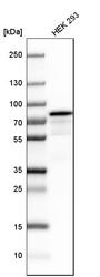
- Experimental details
- Western blot analysis in human cell line HEK 293.
- Sample type
- HUMAN

Supportive validation
- Submitted by
- Atlas Antibodies (provider)
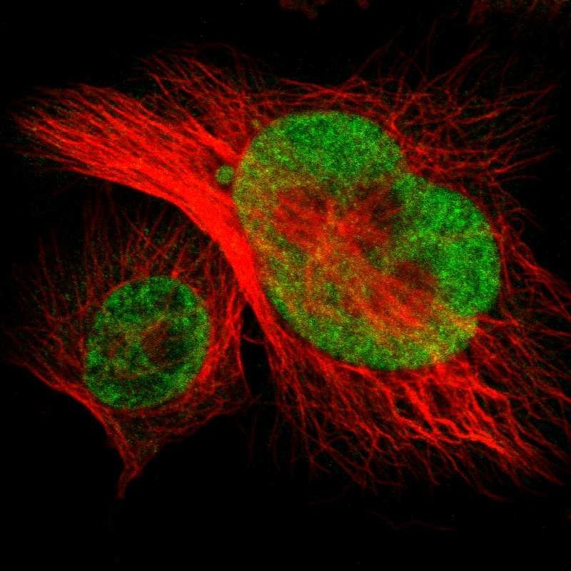
- Main image

- Experimental details
- Immunofluorescent staining of human cell line U-251 MG shows localization to nucleoplasm.
- Sample type
- HUMAN
Enhanced validation
Supportive validation
- Submitted by
- Atlas Antibodies (provider)
- Enhanced method
- Independent antibody validation
- Main image
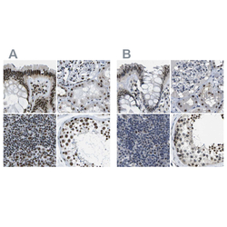
- Experimental details
- Immunohistochemical staining of human colon, kidney, lymph node and testis using Anti-NSRP1 antibody HPA015593 (A) shows similar protein distribution across tissues to independent antibody HPA015603 (B).

Supportive validation
- Submitted by
- Atlas Antibodies (provider)
- Main image

- Experimental details
- Immunohistochemical staining of human gall bladder shows strong nuclear positivity in glandular cells.
- Sample type
- HUMAN

- Submitted by
- Atlas Antibodies (provider)
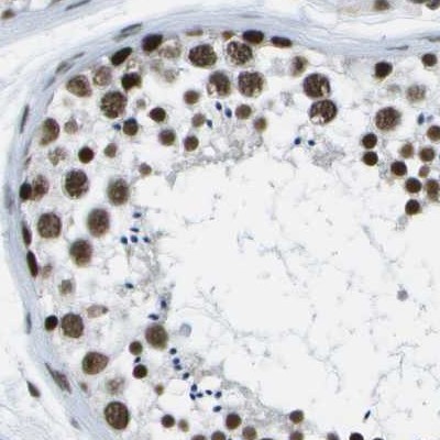
- Main image

- Experimental details
- Immunohistochemical staining of human testis using Anti-NSRP1 antibody HPA015593.
- Sample type
- HUMAN
- Submitted by
- Atlas Antibodies (provider)
- Main image
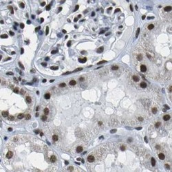
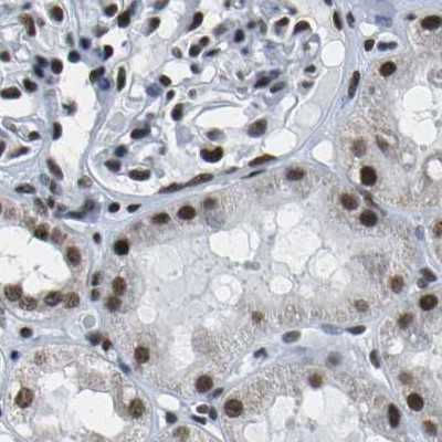
- Experimental details
- Immunohistochemical staining of human kidney using Anti-NSRP1 antibody HPA015593.
- Sample type
- HUMAN
- Submitted by
- Atlas Antibodies (provider)
- Main image

- Experimental details
- Immunohistochemical staining of human lymph node using Anti-NSRP1 antibody HPA015593.
- Sample type
- HUMAN

- Submitted by
- Atlas Antibodies (provider)
- Main image
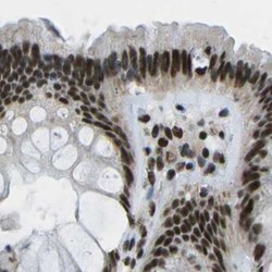
- Experimental details
- Immunohistochemical staining of human colon using Anti-NSRP1 antibody HPA015593.
- Sample type
- HUMAN
