Antibody data
- Antibody Data
- Antigen structure
- References [1]
- Comments [0]
- Validations
- Western blot [1]
- Immunocytochemistry [1]
- Immunohistochemistry [6]
Submit
Validation data
Reference
Comment
Report error
- Product number
- HPA013606 - Provider product page

- Provider
- Atlas Antibodies
- Proper citation
- Atlas Antibodies Cat#HPA013606, RRID:AB_1850717
- Product name
- Anti-HIP1
- Antibody type
- Polyclonal
- Reactivity
- Human, Mouse, Rat
- Host
- Rabbit
- Conjugate
- Unconjugated
- Antigen sequence
NFLRASALSEHISPVVVIPAEASSPDSEPVLEKDD
LMDMDASQQNLFDNKFDDIFGSSFSSDPFNFNSQN
GVNKDEKDHLIERLYREISGLKAQLENMKTESQRV
VLQLKGHVSELEADLAEQQHLRQQAADDCEFLRAE
LDELRRQRED- Isotype
- IgG
- Vial size
- 100 µl
- Storage
- Store at +4°C for short term storage. Long time storage is recommended at -20°C.
Submitted references Clathrin light chains are required for the gyrating-clathrin recycling pathway and thereby promote cell migration.
Majeed SR, Vasudevan L, Chen CY, Luo Y, Torres JA, Evans TM, Sharkey A, Foraker AB, Wong NM, Esk C, Freeman TA, Moffett A, Keen JH, Brodsky FM
Nature communications 2014 May 23;5:3891
Nature communications 2014 May 23;5:3891
No comments: Submit comment
Enhanced validation
- Submitted by
- Atlas Antibodies (provider)
- Enhanced method
- Orthogonal validation
- Main image

- Experimental details
- Western blot analysis in human cell lines A-549 and Caco-2 using Anti-HIP1 antibody. Corresponding HIP1 RNA-seq data are presented for the same cell lines. Loading control: Anti-PPIB.
Supportive validation
- Submitted by
- Atlas Antibodies (provider)
- Main image

- Experimental details
- Immunofluorescent staining of human cell line U-251 MG shows localization to vesicles.
- Sample type
- HUMAN
Enhanced validation
Enhanced validation
Supportive validation
- Submitted by
- Atlas Antibodies (provider)
- Enhanced method
- Orthogonal validation
- Main image

- Experimental details
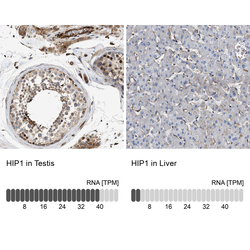
- Immunohistochemistry analysis in human testis and liver tissues using Anti-HIP1 antibody. Corresponding HIP1 RNA-seq data are presented for the same tissues.
- Sample type
- HUMAN
Enhanced validation
- Submitted by
- Atlas Antibodies (provider)
- Enhanced method
- Independent antibody validation
- Main image

- Experimental details
- Immunohistochemical staining of human colon, kidney, liver and testis using Anti-HIP1 antibody HPA013606 (A) shows similar protein distribution across tissues to independent antibody HPA017964 (B).
Supportive validation
- Submitted by
- Atlas Antibodies (provider)
- Main image

- Experimental details
- Immunohistochemical staining of human testis shows high expression.
- Sample type
- HUMAN
- Submitted by
- Atlas Antibodies (provider)
- Main image

- Experimental details
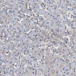
- Immunohistochemical staining of human liver shows low expression as expected.
- Sample type
- HUMAN
- Submitted by
- Atlas Antibodies (provider)
- Main image

- Experimental details
- Immunohistochemical staining of human kidney using Anti-HIP1 antibody HPA013606.
- Sample type
- HUMAN
- Submitted by
- Atlas Antibodies (provider)
- Main image

- Experimental details
- Immunohistochemical staining of human colon using Anti-HIP1 antibody HPA013606.
- Sample type
- HUMAN
 Explore
 Validate
 Learn
Learn Western blot
 Immunocytochemistry