Explore
 Validate
 Learn
Learn Western blot
 Immunocytochemistry
 Immunohistochemistry
Antibody data
- Antibody Data
- Antigen structure
- References [2]
- Comments [0]
- Validations
- Immunocytochemistry [1]
- Immunohistochemistry [6]
Submit
Validation data
Reference
Comment
Report error
- Product number
- HPA013607 - Provider product page

- Provider
- Atlas Antibodies
- Proper citation
- Atlas Antibodies Cat#HPA013607, RRID:AB_1854251
- Product name
- Anti-MYO1B
- Antibody type
- Polyclonal
- Reactivity
- Human
- Host
- Rabbit
- Conjugate
- Unconjugated
- Antigen sequence
LLNKLKLERDFSRYNYLSLDSAKVNGVDDAANFRT
VRNAMQIVGFMDHEAESVLAVVAAVLKLGNIEFKP
ESRVNGLDESKIKDKNELKEICELTGIDQSVLERA
FSFRTVEAKQEKVSTTLNVA- Isotype
- IgG
- Vial size
- 100 µl
- Storage
- Store at +4°C for short term storage. Long time storage is recommended at -20°C.
Submitted references Myosin 1b Regulates Amino Acid Transport by Associating Transporters with the Apical Plasma Membrane of Kidney Cells.
Myosin-II-mediated cell shape changes and cell intercalation contribute to primitive streak formation
Komaba S, Coluccio LM
PloS one 2015;10(9):e0138012
PloS one 2015;10(9):e0138012
Myosin-II-mediated cell shape changes and cell intercalation contribute to primitive streak formation
Rozbicki E, Chuai M, Karjalainen A, Song F, Sang H, Martin R, Knölker H, MacDonald M, Weijer C
Nature Cell Biology 2015 March;17(4):397-408
Nature Cell Biology 2015 March;17(4):397-408
No comments: Submit comment
Supportive validation
- Submitted by
- Atlas Antibodies (provider)
- Main image

- Experimental details
- Immunofluorescent staining of human cell line A-431 shows localization to plasma membrane.
- Sample type
- HUMAN

Enhanced validation
Supportive validation
- Submitted by
- Atlas Antibodies (provider)
- Enhanced method
- Orthogonal validation
- Main image

- Experimental details
- Immunohistochemistry analysis in human liver and pancreas tissues using HPA013607 antibody. Corresponding MYO1B RNA-seq data are presented for the same tissues.
- Sample type
- HUMAN

Supportive validation
- Submitted by
- Atlas Antibodies (provider)
- Main image

- Experimental details
- Immunohistochemical staining of human kidney shows strong cytoplasmic positivity in cells in tubules.

- Submitted by
- Atlas Antibodies (provider)
- Main image
- Experimental details
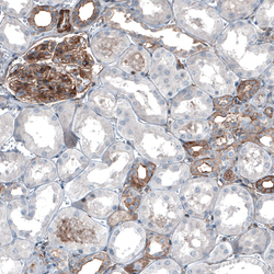
- Immunohistochemical staining of human kidney shows moderate membranous positivity in cells in glomeruli and in cells in tubules.
- Sample type
- HUMAN

- Submitted by
- Atlas Antibodies (provider)
- Main image
- Experimental details
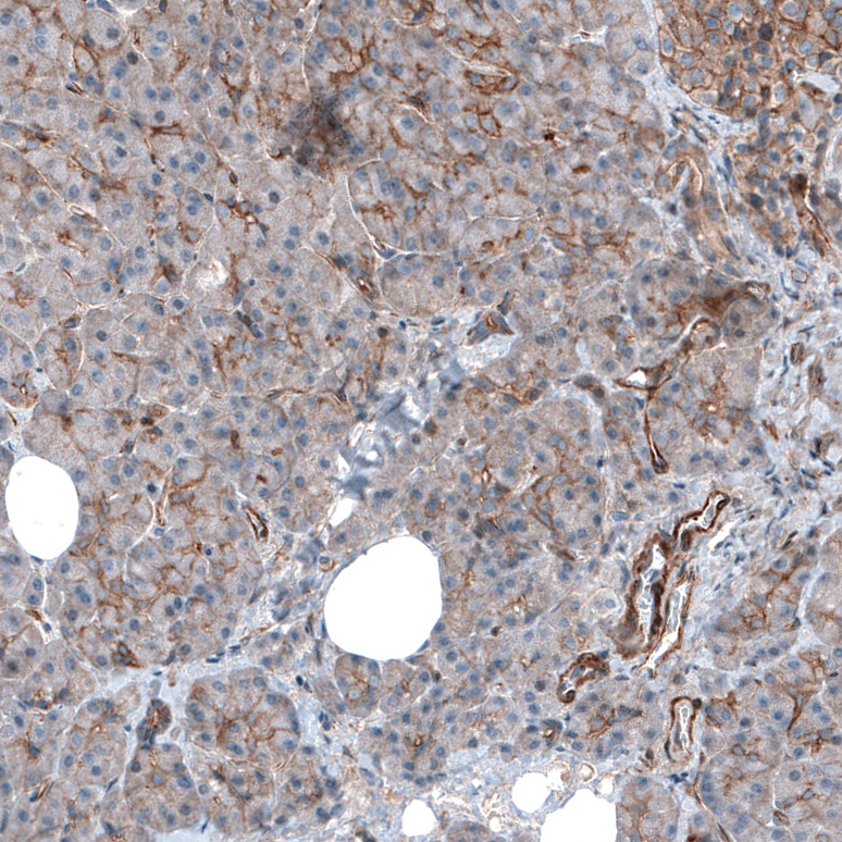
- Immunohistochemical staining of human liver shows moderate membranous positivity in hepatocytes.
- Sample type
- HUMAN

- Submitted by
- Atlas Antibodies (provider)
- Main image

- Experimental details
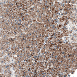
- Immunohistochemical staining of human placenta shows moderate membranous positivity in trophoblastic cells.
- Sample type
- HUMAN

- Submitted by
- Atlas Antibodies (provider)
- Main image

- Experimental details
- Immunohistochemical staining of human pancreas shows moderate membranous positivity in exocrine glandular cells.
- Sample type
- HUMAN