Explore
 Validate
 Learn
Learn Western blot
Antibody data
- Antibody Data
- Antigen structure
- References [0]
- Comments [0]
- Validations
- Western blot [1]
- Immunohistochemistry [1]
Submit
Validation data
Reference
Comment
Report error
- Product number
- ABIN1504531 - Provider product page

- Provider
- antibodies-online
- Product name
- anti-Glycine Receptor, alpha 3 (GLRa3) antibody
- Antibody type
- Polyclonal
- Antigen
- Other
- Description
- Affinity Chromatography
- Reactivity
- Human, Mouse
- Host
- Rabbit
- Isotype
- IgG
- Vial size
- 0.1 mg
- Concentration
- 0.5 mg/mL
No comments: Submit comment
Supportive validation
- Submitted by
- antibodies-online (provider)
- Main image

- Experimental details
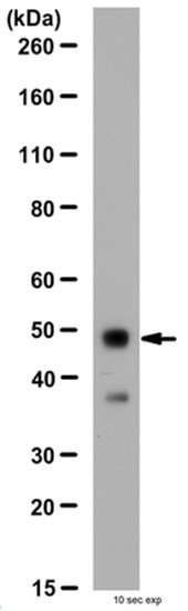
- Western Blot Analysis (Representative lot data):Fetal Human brain lysate was resolved by electrophoresis, transferred to PVDF and probed with anti-Glycine Receptor Alpha 3 subunit. Proteins were visualized using a donkey anti-rabbit secondary antibody conjugated to HRP and a chemiluminescence detection system. Arrow indicates Glycine Receptor ?3 subunit (48kDa).
Supportive validation
- Submitted by
- antibodies-online (provider)
- Main image

- Experimental details
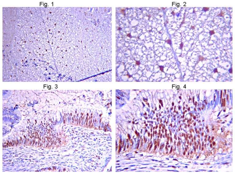
- Immunohistochemistry Analysis: Figures 1+2: Representative positive and background staining patterns on formalin-fixed, paraffin-embedded in Mouse spinal cord. Tissue pretreated with citrate pH 6.0 using antigen retrieval techniques. Antibody diluted to 1/100-1/500 and detected using IHC Select HRP/DAB detection kit protocol. Immunoreactivity is seen as strong staining in Mouse spinal cord. Figure 1 (Low Magnification). Figure 2 (High Magnification). Figures 3 + 4: Representative positive and background staining patterns on formalin-fixed, paraffin-embedded Mouse eye. Tissue pretreated with citrate pH 6.0 using antigen retrieval techniques. Antibody diluted to 1/100-1/300 and detected using IHC Select HRP/DAB detection kit protocol. Immunoreactivity is seen as strong staining in Mouse eye tissue. Figure 3 (Low Magnification). Figure 4 (High Magnification).