Explore
 Validate
 Learn
Learn Immunohistochemistry
Antibody data
- Antibody Data
- Antigen structure
- References [1]
- Comments [0]
- Validations
- Immunohistochemistry [7]
Submit
Validation data
Reference
Comment
Report error
- Product number
- HPA003536 - Provider product page

- Provider
- Atlas Antibodies
- Proper citation
- Atlas Antibodies Cat#HPA003536, RRID:AB_1078936
- Product name
- Anti-LGALS2
- Antibody type
- Polyclonal
- Reactivity
- Human
- Host
- Rabbit
- Conjugate
- Unconjugated
- Antigen sequence
LEVKNMDMKPGSTLKITGSIADGTDGFVINLGQGT
DKLNLHFNPRFSESTIVCNSLDGSNWGQEQREDHL
CFSPGSEVKFTVTFESDKFKVKLPDGHELTFPNRL
GHSHLSYLSVRGGFNMSSFKL- Isotype
- IgG
- Vial size
- 100 µl
- Storage
- Store at +4°C for short term storage. Long time storage is recommended at -20°C.
Submitted references Antibody-based proteomics for discovery and exploration of proteins expressed in pancreatic islets.
Lindskog C, Asplund A, Engkvist M, Uhlen M, Korsgren O, Ponten F
Discovery medicine 2010 Jun;9(49):565-78
Discovery medicine 2010 Jun;9(49):565-78
No comments: Submit comment
Enhanced validation
Supportive validation
- Submitted by
- Atlas Antibodies (provider)
- Enhanced method
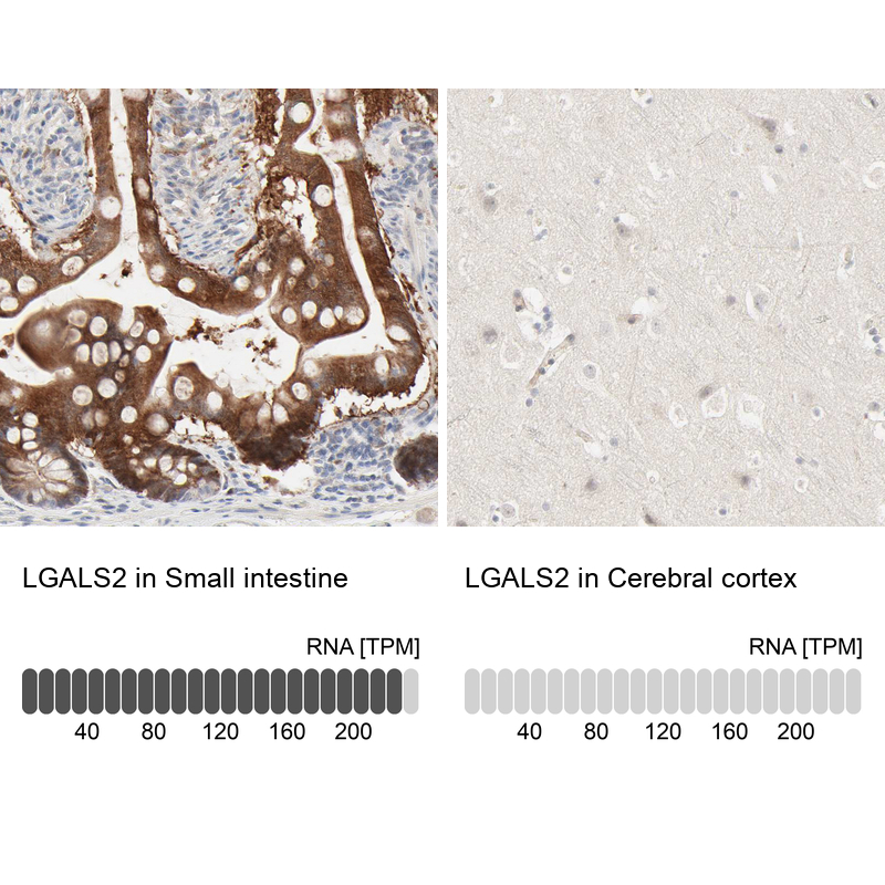
- Orthogonal validation
- Main image

- Experimental details
- Immunohistochemistry analysis in human small intestine and cerebral cortex tissues using HPA003536 antibody. Corresponding LGALS2 RNA-seq data are presented for the same tissues.
- Sample type
- HUMAN
Supportive validation
- Submitted by
- Atlas Antibodies (provider)
- Main image
- Experimental details
- Immunohistochemical staining of human gallbladder shows high expression.
- Sample type
- HUMAN

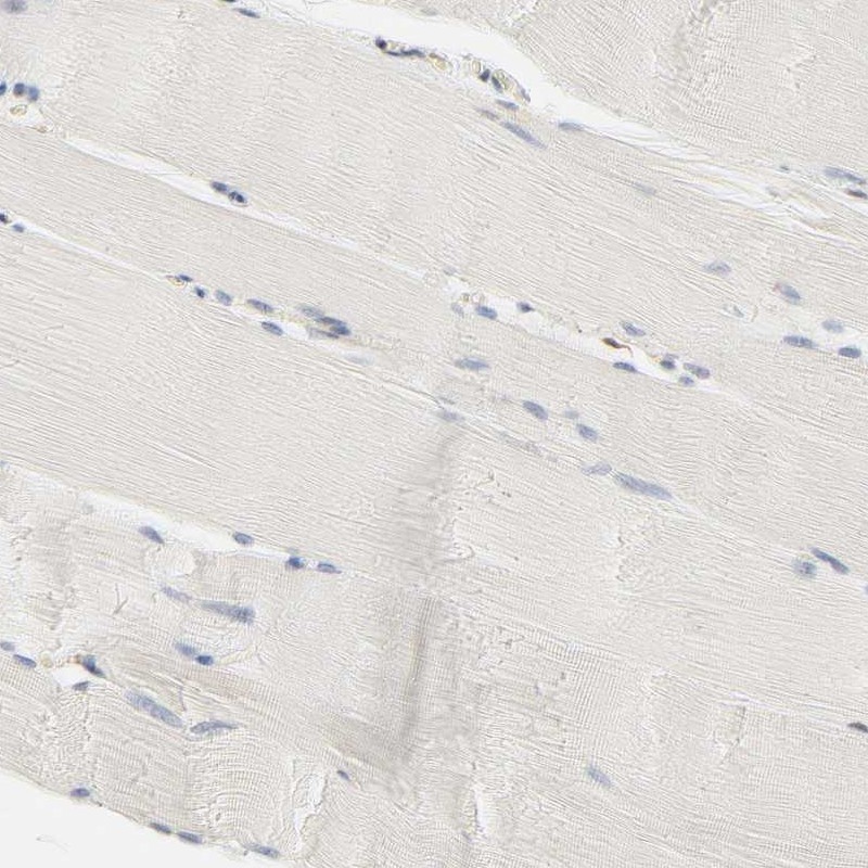
- Submitted by
- Atlas Antibodies (provider)
- Main image
- Experimental details
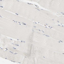
- Immunohistochemical staining of human skeletal muscle shows low expression as expected.
- Sample type
- HUMAN
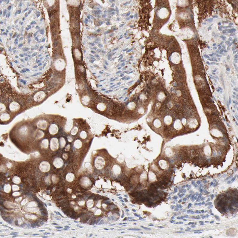
- Submitted by
- Atlas Antibodies (provider)
- Main image

- Experimental details
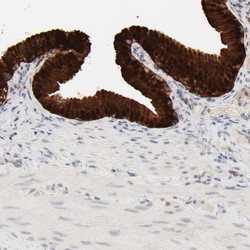
- Immunohistochemical staining of human small intestine shows strong cytoplasmic positivity in glandular cells.
- Sample type
- HUMAN
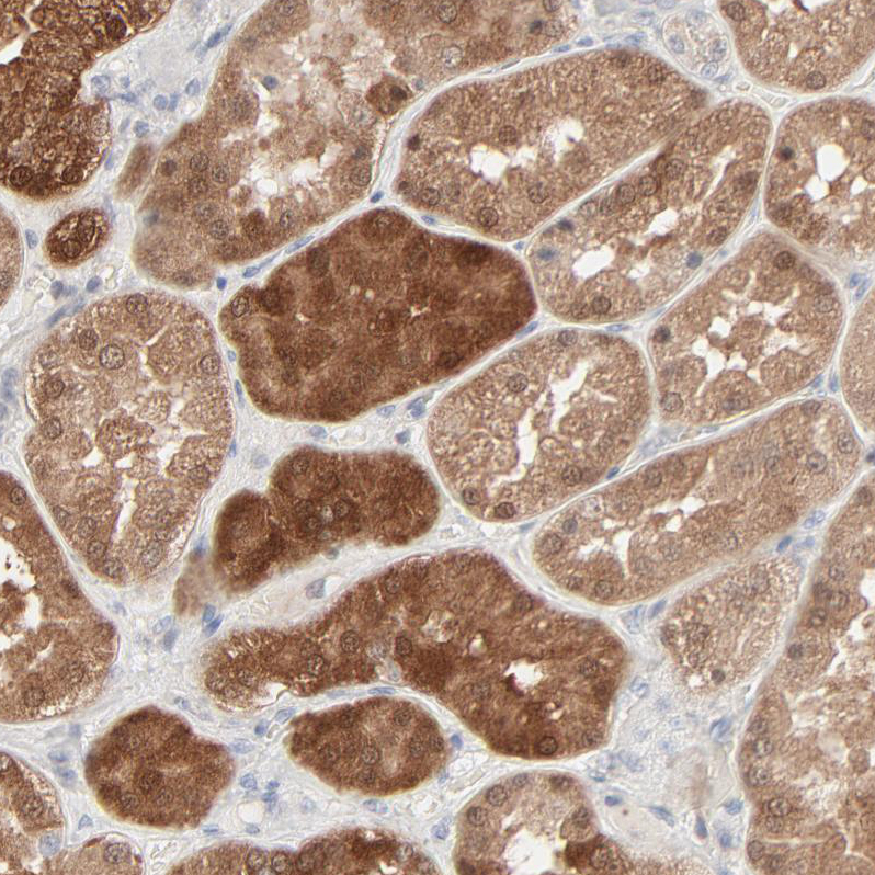
- Submitted by
- Atlas Antibodies (provider)
- Main image
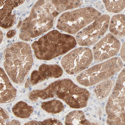
- Experimental details
- Immunohistochemical staining of human kidney shows moderate to strong cytoplasmic positivity in cells in tubules.
- Sample type
- HUMAN
- Submitted by
- Atlas Antibodies (provider)
- Main image

- Experimental details
- Immunohistochemical staining of human lymph node shows strong cytoplasmic positivity in non - germinal center cells.
- Sample type
- HUMAN

- Submitted by
- Atlas Antibodies (provider)
- Main image
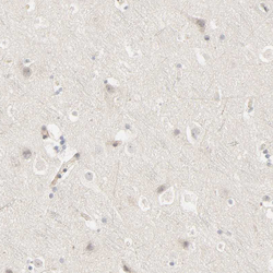
- Experimental details
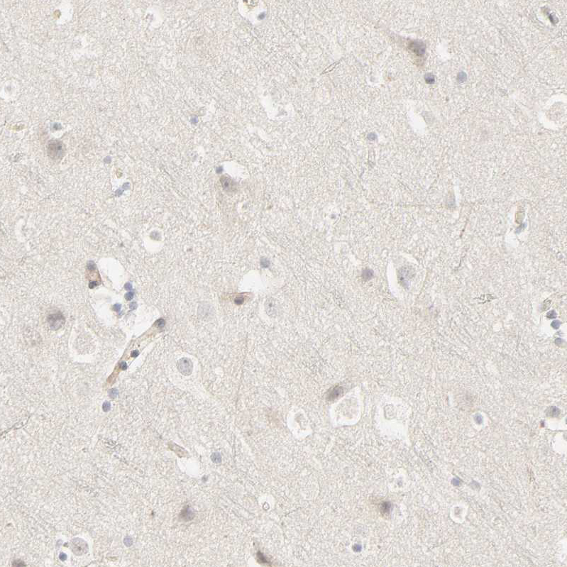
- Immunohistochemical staining of human cerebral cortex shows no positivity in neurons.
- Sample type
- HUMAN