 Explore
 Validate
 Learn
Learn Western blot
Antibody data
- Antibody Data
- Antigen structure
- References [0]
- Comments [0]
- Validations
- Western blot [2]
- Immunocytochemistry [1]
- Flow cytometry [1]
Submit
Validation data
Reference
Comment
Report error
- Product number
- MAB9219-100 - Provider product page

- Provider
- R&D Systems
- Product name
- Human MESP1 Antibody
- Antibody type
- Monoclonal
- Description
- Protein A or G purified from cell culture supernatant. Detects human MESP1 in direct ELISAs.
- Reactivity
- Human
- Host
- Rabbit
- Conjugate
- Unconjugated
- Antigen sequence
Q9BRJ9- Isotype
- IgG
- Antibody clone number
- 2030B
- Vial size
- 100 ug
- Concentration
- 1.0 mg/ml
- Storage
- Use a manual defrost freezer and avoid repeated freeze-thaw cycles. 12 months from date of receipt, -20 to -70 °C as supplied. 1 month, 2 to 8 °C under sterile conditions after reconstitution. 6 months, -20 to -70 °C under sterile conditions after reconstitution.
No comments: Submit comment
Supportive validation
- Submitted by
- R&D Systems (provider)
- Main image
- Experimental details
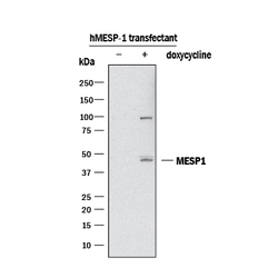
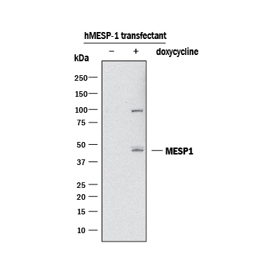
- Detection of Human MESP1 by Western Blot. Western blot shows lysates of Mouse ES cells transfected with human MESP1 untreated (-) or treated (+) with 100 ng/mL Doxycycline overnight. PVDF membrane was probed with 2 µg/mL of Rabbit Anti-Human MESP1 Monoclonal Antibody (Catalog # MAB9219) followed by HRP-conjugated Anti-Rabbit IgG Secondary Antibody (Catalog # HAF008). A specific band was detected for MESP1 at approximately 45 kDa (as indicated). This experiment was conducted under reducing conditions and using Immunoblot Buffer Group 3. Cells provided courtesy of Michael Kyba's lab, University of Minnesota.
- Submitted by
- R&D Systems (provider)
- Main image
- Experimental details
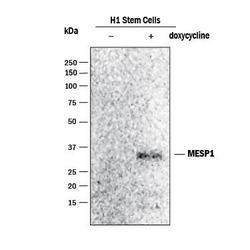
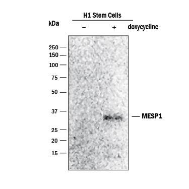
- Detection of Human MESP1 by Western Blot. Western blot shows lysates of H1 human embryonic stem cells untreated (-) or treated (+) with 100 ng/mL Doxycycline overnight. PVDF membrane was probed with Rabbit Anti-Human MESP1 Monoclonal Antibody (Catalog # MAB9219) followed by HRP-conjugated Anti-Rabbit IgG Secondary Antibody (Catalog # HAF008). A specific band was detected for MESP1 at approximately 30 kDa (as indicated). Image provided courtesy of Michael Kyba's lab, University of Minnesota.
Supportive validation
- Submitted by
- R&D Systems (provider)
- Main image
- Experimental details
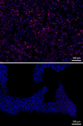
- Human MESP1 in H1 Human Embryonic Stem Cells. MESP1 was detected in fixed H1 human embryonic stem cells untreated (lower panel) or treated (upper panel) with Doxycycline using Rabbit Anti-Human MESP1 Monoclonal Antibody (Catalog # MAB9219). Cells were stained using anti-rabbit IgG secondary antibody (red) and counterstained with DAPI (blue). Image provided courtesy of Michael Kyba's lab, University of Minnesota.

Supportive validation
- Submitted by
- R&D Systems (provider)
- Main image
- Experimental details
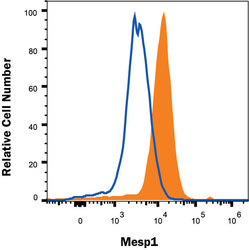
- Detection of MESP1 in Mouse ES cells transfected with human MESP1 by Flow Cytometry. Mouse ES cells transfected with human MESP1 were stained with Rabbit Anti-Human MESP1 Monoclonal Antibody (Catalog # MAB9219, filled histogram) or isotype control antibody (Catalog # MAB1050, open histogram), followed by APC-conjugated Anti-Rabbit IgG Secondary Antibody (Catalog # F0111). To facilitate intracellular staining, cells were fixed and permeabilized with FlowX FoxP3 Fixation & Permeabilization Buffer Kit (Catalog # FC012). View our protocol for Staining Intracellular Molecules.
