Antibody data
- Antibody Data
- Antigen structure
- References [3]
- Comments [0]
- Validations
- Western blot [1]
- Immunocytochemistry [1]
Submit
Validation data
Reference
Comment
Report error
- Product number
- MA1-050 - Provider product page

- Provider
- Invitrogen Antibodies
- Product name
- CETP Monoclonal Antibody (ATM192)
- Antibody type
- Monoclonal
- Antigen
- Synthetic peptide
- Description
- MA1-050 detects cholesteryl ester transfer protein (CETP) from human and rabbit tissues. MA1-050 has been successfully used in Western blot, immunoprecipitation, IF and ELISA protocols. By Western blot, this antibody detects a doublet at ~70 kDa from human serum representing CETP. This antibody can also be used to immunoprecipitate CETP activity from serum samples using protein G. The MA1-050 immunogen is a synthetic peptide corresponding to residues C D(149) S G R V R T D A P D(159) of human CETP protein.
- Reactivity
- Human, Rabbit
- Host
- Mouse
- Isotype
- IgG
- Antibody clone number
- ATM192
- Vial size
- 100 µL
- Concentration
- 2 mg/mL
- Storage
- -20° C, Avoid Freeze/Thaw Cycles
Submitted references Cholesteryl Ester Transfer Protein Inhibition With Anacetrapib Decreases Fractional Clearance Rates of High-Density Lipoprotein Apolipoprotein A-I and Plasma Cholesteryl Ester Transfer Protein.
Mouse monoclonal antipeptide antibodies specific for cholesteryl ester transfer protein (CETP).
Mouse monoclonal antipeptide antibodies specific for cholesteryl ester transfer protein (CETP).
Reyes-Soffer G, Millar JS, Ngai C, Jumes P, Coromilas E, Asztalos B, Johnson-Levonas AO, Wagner JA, Donovan DS, Karmally W, Ramakrishnan R, Holleran S, Thomas T, Dunbar RL, deGoma EM, Rafeek H, Baer AL, Liu Y, Lassman ME, Gutstein DE, Rader DJ, Ginsberg HN
Arteriosclerosis, thrombosis, and vascular biology 2016 May;36(5):994-1002
Arteriosclerosis, thrombosis, and vascular biology 2016 May;36(5):994-1002
Mouse monoclonal antipeptide antibodies specific for cholesteryl ester transfer protein (CETP).
Thomas AP, Smith AM, Cumming RI, Jones C, Thomas RC, Pleasants KT, Barakat H
Hybridoma 1996 Oct;15(5):359-64
Hybridoma 1996 Oct;15(5):359-64
Mouse monoclonal antipeptide antibodies specific for cholesteryl ester transfer protein (CETP).
Thomas AP, Smith AM, Cumming RI, Jones C, Thomas RC, Pleasants KT, Barakat H
Hybridoma 1996 Oct;15(5):359-64
Hybridoma 1996 Oct;15(5):359-64
No comments: Submit comment
Supportive validation
- Submitted by
- Invitrogen Antibodies (provider)
- Main image

- Experimental details
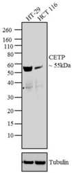
- Western blot analysis was performed on whole cell extracts of HT-29 (Lane 1) and HCT 116 (Lane 2). The blot was probed with Anti-CETP Mouse Monoclonal Antibody (Product # MA1-050, 2 µg/mL) and detected by chemiluminescence using Goat anti-Mouse IgG (H+L) Superclonal™ Secondary Antibody, HRP conjµgate (Product # A28177, 0.4 µg/mL, 1:2500 dilution). A ~ 55 kDa band corresponding to CETP was observed in the cell lines tested. Known quantity of protein samples were electrophoresed using Novex® NuPAGE® 4-12 % Bis-Tris gel (Product # NP0321BOX), XCell SureLock™ Electrophoresis System (Product # EI0002) and Novex® Sharp Pre-Stained Protein Standard (Product # LC5800). Resolved proteins were then transferred onto a nitrocellulose membrane with iBlot® 2 Dry Blotting System (Product # IB21001). The membrane was probed with the relevant primary and secondary Antibody following blocking with 5 % skimmed milk. Chemiluminescent detection was performed using Pierce™ ECL Western Blotting Substrate (Product # 32106).
Supportive validation
- Submitted by
- Invitrogen Antibodies (provider)
- Main image

- Experimental details
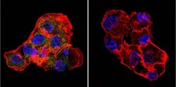
- Immunofluorescent analysis of CETP in HepG2 Cells. Cells were grown on chamber slides and fixed with formaldehyde prior to staining. Cells were probed without (control) or with a CETP monoclonal antibody (Product # MA1-050) at a dilution of 1:20 overnight at 4 C, washed with PBS and incubated with a DyLight-488 conjugated secondary antibody (Product # 35503). CETP staining (green), F-Actin staining with Phalloidin (red) and nuclei with DAPI (blue) is shown. Images were taken at 60X magnification.
 Explore
 Validate
 Learn
Learn Western blot
 ELISA
ELISA