Explore
 Validate
 Learn
LearnPA5-29092
antibody from Invitrogen Antibodies
Targeting: TRIOBP
DFNB28, HRIHFB2122, KIAA1662, TAP68, Tara
 Western blot
Antibody data
- Antibody Data
- Antigen structure
- References [1]
- Comments [0]
- Validations
- Western blot [4]
- Immunocytochemistry [1]
- Immunohistochemistry [1]
- Other assay [1]
Submit
Validation data
Reference
Comment
Report error
- Product number
- PA5-29092 - Provider product page

- Provider
- Invitrogen Antibodies
- Product name
- Tara Polyclonal Antibody
- Antibody type
- Polyclonal
- Antigen
- Recombinant protein fragment
- Description
- Recommended positive controls: 293T, A431, HeLa, HepG2, Rat2.
- Concentration
- 1 mg/mL
Submitted references Regulation of the actin cytoskeleton by the Ndel1-Tara complex is critical for cell migration.
Hong JH, Kwak Y, Woo Y, Park C, Lee SA, Lee H, Park SJ, Suh Y, Suh BK, Goo BS, Mun DJ, Sanada K, Nguyen MD, Park SK
Scientific reports 2016 Aug 22;6:31827
Scientific reports 2016 Aug 22;6:31827
No comments: Submit comment
Supportive validation
- Submitted by
- Invitrogen Antibodies (provider)
- Main image

- Experimental details
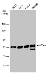
- Western blot analysis of Tara using 30 µg of A) HeLa and B) HepG2 lysate. Samples were loaded onto a 10% SDS-PAGE gel and probed with a Tara polyclonal antibody (Product # PA5-29092) at a dilution of 1:1000.

- Submitted by
- Invitrogen Antibodies (provider)
- Main image
- Experimental details
- Western blot analysis of Tara using 30 µg of NIH-3T3 lysate. Samples were loaded onto a 10% SDS-PAGE gel and probed with a Tara polyclonal antibody (Product # PA5-29092) at a dilution of 1:1000.

- Submitted by
- Invitrogen Antibodies (provider)
- Main image
- Experimental details
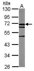
- Western Blot analysis of Tara was performed by separating 30 µg of various whole cell extracts by 7.5% SDS-PAGE. Proteins were transferred to a membrane and probed with a Tara Polyclonal Antibody (Product # PA5-29092) at a dilution of 1:1000 and a HRP-conjugated anti-rabbit IgG secondary antibody.

- Submitted by
- Invitrogen Antibodies (provider)
- Main image

- Experimental details
- Western Blot analysis of Tara was performed by separating 30 µg of Whole cell extracts by 7.5% SDS-PAGE. Proteins were transferred to a membrane and probed with a Tara Polyclonal Antibody (Product # PA5-29092) at a dilution of 1:5000. The HRP-conjugated anti-rabbit IgG antibody was used to detect the primary antibody.

Supportive validation
- Submitted by
- Invitrogen Antibodies (provider)
- Main image

- Experimental details
- Immunofluorescent analysis of Tara in paraformaldehyde-fixed HeLa cells using a Tara polyclonal antibody (Product # PA5-29092) at a 1:200 dilution.

Supportive validation
- Submitted by
- Invitrogen Antibodies (provider)
- Main image

- Experimental details
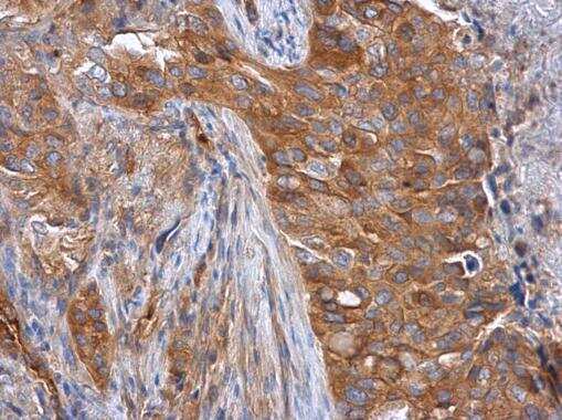
- Immunohistochemistry (Paraffin) analysis of Tara was performed in paraffin-embedded human lung cancer tissue using Tara Polyclonal Antibody (Product # PA5-29092) at a dilution of 1:500.
Supportive validation
- Submitted by
- Invitrogen Antibodies (provider)
- Main image

- Experimental details
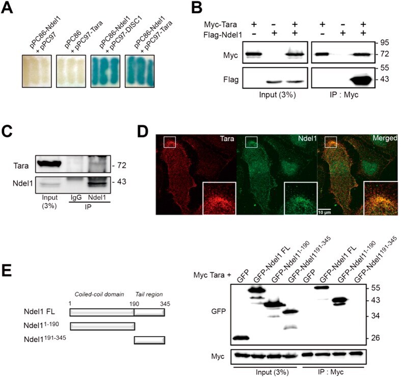
- Figure 1 Tara interacts with Ndel1. ( A ) Interactions of Tara and Ndel1 in a yeast two-hybrid assay. Interaction-dependent beta-galactosidase expression of co-transformants with the indicated constructs are shown. Co-transfection with pPC97-DISC1 and pPC86-Ndel1 was used as a positive control. ( B ) Co-immunoprecipitation of Ndel1 with full-length Tara. Flag-Ndel1 and Myc-Tara were transiently transfected into HEK293 cells and lysates were immunoprecipitated with anti-Myc. Immunoprecipitates were analyzed by immunoblotting with anti-Flag and anti-Myc. IP, immunoprecipitation. ( C ) Co-immunoprecipitation of endogenous Tara and Ndel1 from HEK293 cell lysates. Anti-Ndel1 immunoprecipitates were analyzed by immunoblotting with anti-Ndel1 and anti-Tara antibodies. ( D ) Ndel1 co-localizes with Tara at the peripheral region of SH-SY5Y cells. Endogenous Tara and Ndel1 were stained with anti-Tara (red) and anti-Ndel1 antibody (green), respectively. ( E ) HEK293 cell lysates expressing Myc-Tara and GFP-tagged fragments of Ndel1 were immunoprecipitated with anti-Myc antibody and immunoblots were probed with anti-Myc and anti-GFP.