Explore
 Validate
 Learn
Learn Western blot
 ELISA
ELISAAntibody data
- Antibody Data
- Antigen structure
- References [3]
- Comments [0]
- Validations
- Western blot [1]
- Immunocytochemistry [1]
Submit
Validation data
Reference
Comment
Report error
- Product number
- 21733-1-AP - Provider product page

- Provider
- Proteintech Group
- Proper citation
- Proteintech Cat#21733-1-AP, RRID:AB_10732728
- Product name
- KANK2 antibody
- Antibody type
- Polyclonal
- Description
- KANK2 antibody (Cat. #21733-1-AP) is a rabbit polyclonal antibody that shows reactivity with human, mouse, rat and has been validated for the following applications: IF, WB, ELISA.
- Reactivity
- Human, Mouse, Rat
- Host
- Rabbit
- Conjugate
- Unconjugated
- Isotype
- IgG
- Vial size
- 20ul, 150ul
Submitted references Nephrotic-syndrome-associated mutation of KANK2 induces pathologic binding competition with physiological interactor KIF21A.
Knockout of the neonatal Fc receptor in cultured podocytes alters IL-6 signaling and the actin cytoskeleton.
The RNA-binding protein Fus directs translation of localized mRNAs in APC-RNP granules.
Xu Y, Guo C, Pan W, Zhao C, Ding Y, Xie X, Wei Z, Sun Y, Yu C
The Journal of biological chemistry 2021 Aug;297(2):100958
The Journal of biological chemistry 2021 Aug;297(2):100958
Knockout of the neonatal Fc receptor in cultured podocytes alters IL-6 signaling and the actin cytoskeleton.
Tonsawan P, Dylewski J, Lewis L, Blaine J
American journal of physiology. Cell physiology 2019 Nov 1;317(5):C1048-C1060
American journal of physiology. Cell physiology 2019 Nov 1;317(5):C1048-C1060
The RNA-binding protein Fus directs translation of localized mRNAs in APC-RNP granules.
Yasuda K, Zhang H, Loiselle D, Haystead T, Macara IG, Mili S
The Journal of cell biology 2013 Dec 9;203(5):737-46
The Journal of cell biology 2013 Dec 9;203(5):737-46
No comments: Submit comment
Supportive validation
- Submitted by
- Proteintech Group (provider)
- Main image

- Experimental details
- mouse lung tissue were subjected to SDS PAGE followed by western blot with 21733-1-AP(KANK2 antibody) at dilution of 1:800
- Sample type
- tissue

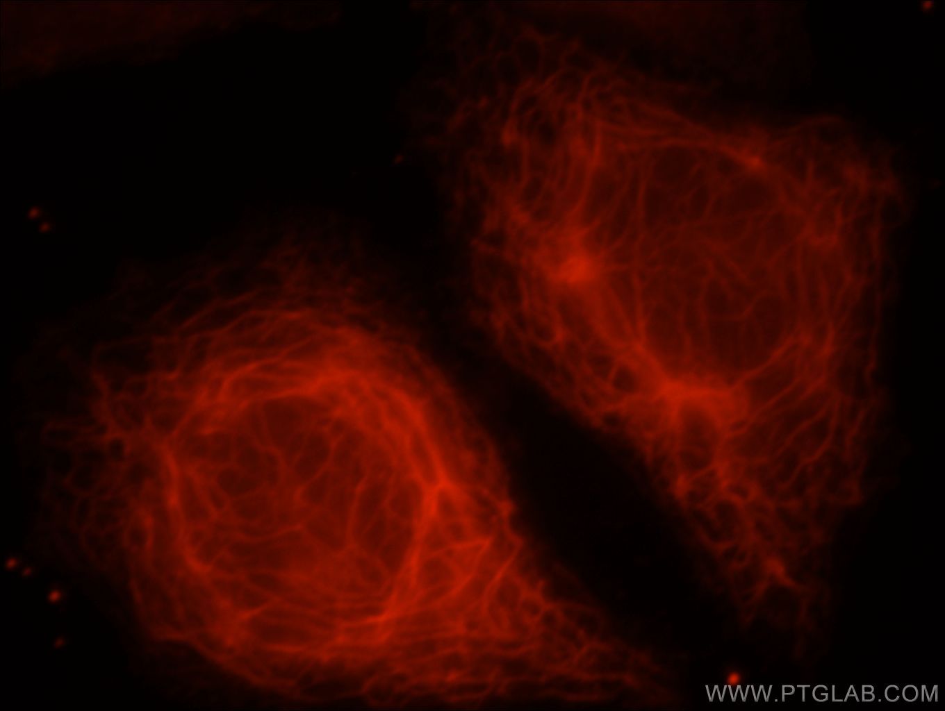
Supportive validation
- Submitted by
- Proteintech Group (provider)
- Main image

- Experimental details
- Immunofluorescent analysis of HepG2 cells, using KANK2 antibody 21733-1-AP at 1:25 dilution and Rhodamine-labeled goat anti-rabbit IgG (red).
- Sample type
- cell line