Antibody data
- Antibody Data
- Antigen structure
- References [1]
- Comments [0]
- Validations
- Western blot [1]
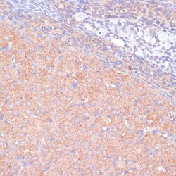
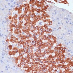
- Immunohistochemistry [2]
- Other assay [3]
Submit
Validation data
Reference
Comment
Report error
- Product number
- PA5-116620 - Provider product page

- Provider
- Invitrogen Antibodies
- Product name
- KANK2 Polyclonal Antibody
- Antibody type
- Polyclonal
- Antigen
- Recombinant full-length protein
- Description
- Positive Samples: Mouse skeletal muscle, Mouse heart, Mouse kidney Immunogen sequence: MAQVLHVPAP FPGTPGPASP PAFPAKDPDP PYSVETPYGY RLDLDFLKYV DDIEKGHTLR RVAVQRRPRL SSLPRGPGSW WTSTESLCSN ASGDSRHSAY SYCGRGFYPQ YGALETRGGF NPRVERTLLD ARRRLEDQAA TPTGLGSLTP SAAGSTASLV GVGLPPPTPR SSGLSTPVPP SAGHLAHVRE QMAGALRKLR QLEEQVKLIP VLQVKLSVLQ EEKRQLTVQL KSQKFLGHPT AGRGRSELCL DLPDPPEDPV ALETRSVGTW VRERDLGMPD GEAALAAKVA VLETQLKKAL
- Reactivity
- Human, Mouse, Rat
- Host
- Rabbit
- Isotype
- IgG
- Vial size
- 100 µL
- Concentration
- 1.33 mg/mL
- Storage
- -20° C, Avoid Freeze/Thaw Cycles
Submitted references HSP70 Ameliorates Septic Lung Injury via Inhibition of Apoptosis by Interacting with KANK2.
Pei Q, Ni W, Yuan Y, Yuan J, Zhang X, Yao M
Biomolecules 2022 Mar 7;12(3)
Biomolecules 2022 Mar 7;12(3)
No comments: Submit comment
Supportive validation
- Submitted by
- Invitrogen Antibodies (provider)
- Main image

- Experimental details
- Western blot analysis of KANK2 using KANK2 Polyclonal Antibody (Product # PA5-116620) at a 1:1,000 dilution. Secondary antibody: HRP Goat Anti-Rabbit IgG (H+L) at 1:10,000 dilution. Lysates/proteins: 25ug per lane. Blocking buffer: 3% nonfat dry milk in TBST.
Supportive validation
- Submitted by
- Invitrogen Antibodies (provider)
- Main image

- Experimental details
- Immunohistochemistry (Paraffin) analysis of KANK2 in rat ovary tissue using KANK2 Polyclonal Antibody (Product # PA5-116620) at a dilution of 1:100.
- Submitted by
- Invitrogen Antibodies (provider)
- Main image

- Experimental details
- Immunohistochemistry (Paraffin) analysis of KANK2 in rat pancreas tissue using KANK2 Polyclonal Antibody (Product # PA5-116620) at a dilution of 1:100.
Supportive validation
- Submitted by
- Invitrogen Antibodies (provider)
- Main image

- Experimental details
- Figure 4 RNA sequencing and proteomics in HEK 293T cells and validation in A549 cells. HEK 293T cells were transfected with hsp70 -plasmid. Transcriptomes analysis was conducted in HEK 293T cells by RNA sequencing. Heat map of the cluster analysis ( A ) and volcano plot ( B ) was presented. The protein binding to HSP70 was extracted by IP (anti-Flag-HSP70). Then, the protein was separated by electrophoresis and stained with silver ( C ). Proteomics analysis was conducted by MS. Enrichment analysis for the binding protein was shown ( D ). IP was performed with an anti-Flag antibody. The expressions of HA-KANK2 and Flag-HSP70 were measured in HEK 293Ts. KANK2 bands were indicated with black arrow ( E ). A549 cells were cultured. The mRNA ( F ) and protein ( G ) levels of KANK2 in HSP OE A549 cells were measured using qPCR and WB. IP was performed with an anti-HSP70 antibody, and KANK2 was assessed in A549 cells ( H ). Representative immunofluorescence images of HSP70 and KANK2 in the A549 cells were acquired ( I ). Bar = 10 um. BP, biological process; CC, cellular component; MF, molecular function; MS, mass spectrometry. ** p < 0.01.
- Submitted by
- Invitrogen Antibodies (provider)
- Main image

- Experimental details
- Interaction of HSP70 and KANK2 in A549 cells. WT and HSP70 OE A549 cells were treated with LPS (4 ug/mL) and si-KANK2 (50 nM, si-NC was set as corresponding control). Cell viability ( A ) was measured by CCK-8 and cell colony formation ( B , G ) was evaluated by crystal violet staining. Dead cells were enumerated by flow cytometry labelling with Annexin V and PI ( C , H ). The levels of KANK2 and released AIF were measured using WB ( D , E ). Mitochondrial membrane permeability was assessed by mito-tracker and fluorescence intensity was measured by imaging system ( F ). Data are presented as mean +- SD of data from three independent experiments. Bar = 200 um. ** p < 0.01.
- Submitted by
- Invitrogen Antibodies (provider)
- Main image

- Experimental details
- Interaction of HSP70 and KANK2 in hsp70.1 -/- mice. hsp70.1 +/+ , hsp70.1 +/- , and hsp70.1 -/- mice were bred and the lung tissues were collected. The levels of HSP70 were measured in mice lung by WB ( A ). Gross profile of mice before and after CLP was assessed ( B ). The pathological appearances of lung tissue were evaluated by H&E staining ( C , E ). The KANK2-positive cells were evaluated by IHC. Brown color indicates positive staining of KANK2 ( D , F ). The levels of HSP70, KANK2, and released AIF were measured using WB ( G ). Data are presented as mean +- SD of data from three independent experiments. ** p < 0.01.
 Explore
 Validate
 Learn
Learn Western blot