 Explore
 Validate
 Learn
Learn Western blot
Antibody data
- Antibody Data
- Antigen structure
- References [0]
- Comments [0]
- Validations
- Western blot [3]
- Immunocytochemistry [2]
- Immunohistochemistry [3]
Submit
Validation data
Reference
Comment
Report error
- Product number
- GTX105295 - Provider product page

- Provider
- GeneTex
- Proper citation
- GeneTex Cat#GTX105295, RRID:AB_1951348
- Product name
- PPP1CB antibody
- Antibody type
- Polyclonal
- Reactivity
- Human, Mouse
- Host
- Rabbit
No comments: Submit comment
Supportive validation
- Submitted by
- GeneTex (provider)
- Main image

- Experimental details
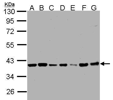
- Sample (30 ?g whole cell lysate)A: 293TB: A431 (GTX27909)C: H1299D: HeLa S3 (GTX14654)E: HepG2 (GTX27900)F: MOLT4 (GTX27912)G: Raji (GTX27908) 7.5% SDS PAGEGTX105295 diluted at 1:1000The HRP-conjugated anti-rabbit IgG antibody (GTX213110-01) was used to detect the primary antibody.
- Submitted by
- GeneTex (provider)
- Main image

- Experimental details
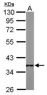
- Sample (50 ?g of whole cell lysate) A: Mouse brain 10% SDS PAGE GTX105295 diluted at 1:1000 The HRP-conjugated anti-rabbit IgG antibody (GTX213110-01) was used to detect the primary antibody.
- Submitted by
- GeneTex (provider)
- Main image
- Experimental details
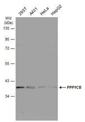
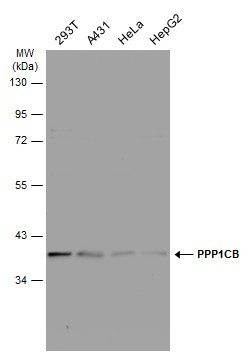
- Various whole cell extracts (30 ?g) were separated by 10% SDS-PAGE, and the membrane was blotted with PPP1CB antibody (GTX105295) diluted at 1:1000. The HRP-conjugated anti-rabbit IgG antibody (GTX213110-01) was used to detect the primary antibody.
Supportive validation
- Submitted by
- GeneTex (provider)
- Main image
- Experimental details
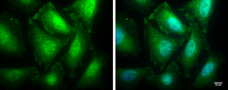
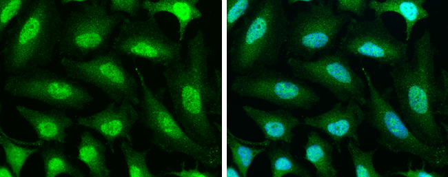
- PPP1CB antibody detects PPP1CB protein at cytoplasm and nucleus by immunofluorescent analysis.Sample: HeLa cells were fixed in 4% paraformaldehyde for 10 min.Green: PPP1CB protein stained by PPP1CB antibody (GTX105295) diluted at 1:100.Blue: Hoechst 33342 staining.Scale bar = 10 £gm.
- Submitted by
- GeneTex (provider)
- Main image

- Experimental details
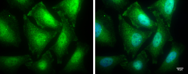
- PPP1CB antibody detects PPP1CB protein at cytoplasm and nucleus by immunofluorescent analysis.Sample: HeLa cells were fixed in 4% paraformaldehyde at RT for 15 min.Green: PPP1CB stained by PPP1CB antibody (GTX105295) diluted at 1:500.Blue: Hoechst 33342 staining.
Supportive validation
- Submitted by
- GeneTex (provider)
- Main image
- Experimental details
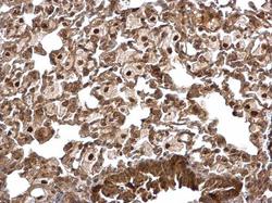
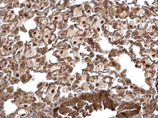
- PPP1CB antibody detects PPP1CB protein at nucleus and cytosol on mouse lung by immunohistochemical analysis. Sample: Paraffin-embedded mouse lung. PPP1CB antibody (GTX105295) dilution: 1:500.
- Submitted by
- GeneTex (provider)
- Main image

- Experimental details
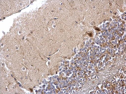
- PPP1CB antibody detects PPP1CB protein at nucleus and cytosol on mouse hind brain by immunohistochemical analysis. Sample: Paraffin-embedded mouse hind brain. PPP1CB antibody (GTX105295) dilution: 1:500.
- Submitted by
- GeneTex (provider)
- Main image
- Experimental details
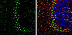
- PPP1CB antibody detects PPP1CB Protein expression by immunohistochemical analysis.Sample: Frozen-sectioned adult mouse cerebellum. Green: PPP1CB stained by PPP1CB antibody (GTX105295) diluted at 1:250.Red: NF-H, stained by NF-H antibody [GT114] (GTX634289) diluted at 1:500.Blue: Fluoroshield with DAPI (GTX30920).
