Explore
 Validate
 Learn
Learn Western blot
Antibody data
- Antibody Data
- Antigen structure
- References [1]
- Comments [0]
- Validations
- Western blot [2]
- Immunocytochemistry [2]
- Immunohistochemistry [1]
Submit
Validation data
Reference
Comment
Report error
- Product number
- PA5-30098 - Provider product page

- Provider
- Invitrogen Antibodies
- Product name
- NCBP1 Polyclonal Antibody
- Antibody type
- Polyclonal
- Antigen
- Recombinant protein fragment
- Description
- Recommended positive controls: A549, HeLa, HepG2.
- Concentration
- 1 mg/mL
Submitted references mRNA export through an additional cap-binding complex consisting of NCBP1 and NCBP3.
Gebhardt A, Habjan M, Benda C, Meiler A, Haas DA, Hein MY, Mann A, Mann M, Habermann B, Pichlmair A
Nature communications 2015 Sep 18;6:8192
Nature communications 2015 Sep 18;6:8192
No comments: Submit comment
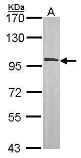
Supportive validation
- Submitted by
- Invitrogen Antibodies (provider)
- Main image
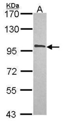
- Experimental details
- Western Blot using NCBP1 Polyclonal Antibody (Product # PA5-30098). Sample (30 µg of whole cell lysate). Lane A: Hela . 7.5% SDS PAGE. NCBP1 Polyclonal Antibody (Product # PA5-30098) diluted at 1:1,000.
- Submitted by
- Invitrogen Antibodies (provider)
- Main image
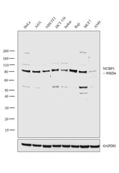
- Experimental details
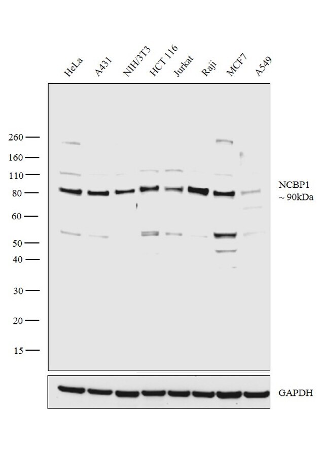
- Western blot analysis was performed on whole cell extracts (30 µg lysate) of HeLa (Lane 1), A431 (Lane 2), NIH/3T3 (Lane 3), HCT 116 (Lane 4), Jurkat (Lane 5), Raji (Lane 6), MCF7 (Lane 7) and A549 (Lane 8). The blot was probed with Anti-NCBP1 Polyclonal Antibody (Product #PA5-30098, 1:2000 dilution) and detected by chemiluminescence using Goat anti-Rabbit IgG (H+L) Superclonal™ Secondary Antibody, HRP conjugate (Product # A27036, 0.25 µg/ml, 1:4000 dilution). A 90 kDa band corresponding to NCBP1 was observed in all the cell lines tested.
Supportive validation
- Submitted by
- Invitrogen Antibodies (provider)
- Main image
- Experimental details
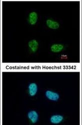
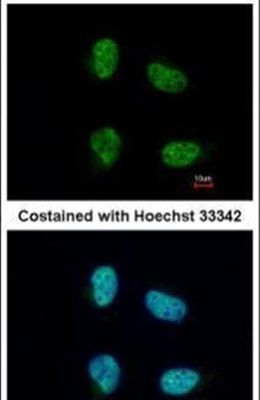
- Immunofluorescent analysis of CBP80 in paraformaldehyde-fixed HeLa cells using a CBP80 polyclonal antibody (Product # PA5-30098) at a 1:200 dilution.
- Submitted by
- Invitrogen Antibodies (provider)
- Main image
- Experimental details
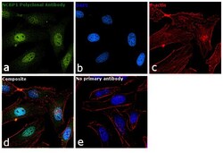
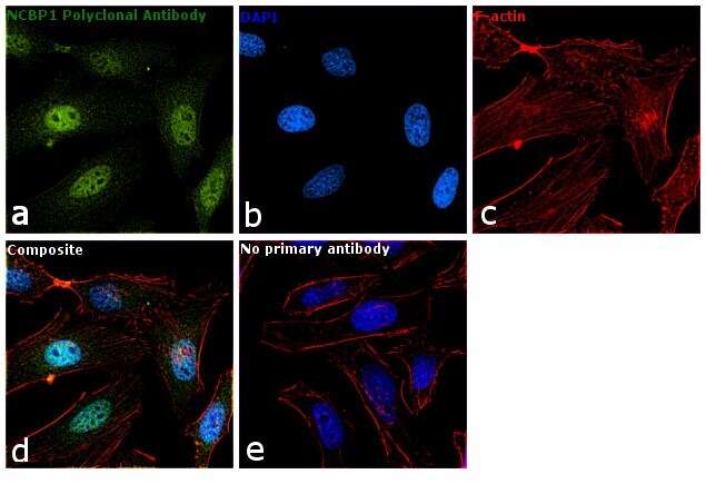
- Immunofluorescence analysis of NCBP1 was performed using 70% confluent log phase HeLa cells. The cells were fixed with 4% paraformaldehyde for 10 minutes, permeabilized with 0.1% Triton™ X-100 for 15 minutes, and blocked with 1% BSA for 1 hour at room temperature. The cells were labeled with NCBP1 Polyclonal Antibody (Product # PA5-30098) at 1:100 dilution in 0.1% BSA, incubated at 4 degree Celsius overnight and then labeled with Goat anti-Rabbit IgG (H+L) Superclonal™ Secondary Antibody, Alexa Fluor® 488 conjugate (Product # A27034) at a dilution of 1:2000 for 45 minutes at room temperature (Panel a: green). Nuclei (Panel b: blue) were stained with ProLong™ Diamond Antifade Mountant with DAPI (Product # P36962). F-actin (Panel c: red) was stained with Rhodamine Phalloidin (Product # R415). Panel d represents the merged image showing predominant nucleus and cytoplasmic localization. Panel e represents control cells with no primary antibody to assess background. The images were captured at 60X magnification.
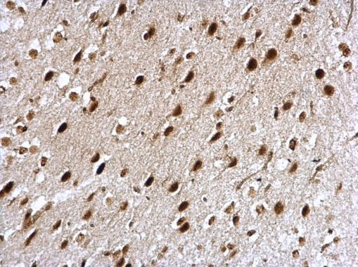
Supportive validation
- Submitted by
- Invitrogen Antibodies (provider)
- Main image

- Experimental details
- NCBP1 Polyclonal Antibody detects CBP80 protein at nucleus on mouse fore brain by immunohistochemical analysis. Sample: Paraffin-embedded mouse fore brain. NCBP1 Polyclonal Antibody (Product # PA5-30098) dilution: 1:500. Antigen Retrieval: EDTA based buffer, pH 8.0, 15 min.