Explore
 Validate
 Learn
Learn Western blot
Antibody data
- Antibody Data
- Antigen structure
- References [0]
- Comments [0]
- Validations
- Western blot [2]
- ELISA [1]
- Immunocytochemistry [1]
- Immunohistochemistry [1]
Submit
Validation data
Reference
Comment
Report error
- Product number
- TA590762 - Provider product page

- Provider
- OriGene
- Product name
- Rabbit Polyclonal cleavage stimulation factor Antibody
- Antibody type
- Polyclonal
- Description
- Rabbit Polyclonal cleavage stimulation factor Antibody
- Host
- Rabbit
- Conjugate
- Unconjugated
- Epitope
- CSTF1
- Isotype
- IgG
- Antibody clone number
- NULL
- Vial size
- 100 µg
- Concentration
- 1.04mg/ml
No comments: Submit comment
Supportive validation
- Submitted by
- OriGene (provider)
- Main image

- Experimental details
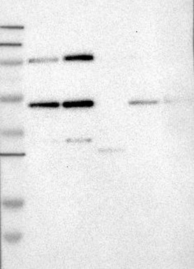
- Lane 1: Marker [kDa] 250, 130, 95, 72, 55, 36, 28, 17, 11; Lane 2: RT-4; Lane 3: U-251 MG; Lane 4: Human Plasma; Lane 5: Liver; Lane 6: TonsilThis validation was performed by Protein Atlas and the presentation of data is for informational purposes only.
- Validation comment
- WB
- Submitted by
- OriGene (provider)
- Main image

- Experimental details
- HEK293T cells were transfected with the pCMV6-ENTRY control (Left lane) or pCMV6-ENTRY CSTF1 (RC219441, Right lane) cDNA for 48 hrs and lysed. Equivalent amounts of cell lysates (5 ug per lane) were separated by SDS-PAGE and immunoblotted with anti-CSTF1.
- Validation comment
- WB

Supportive validation
- Submitted by
- OriGene (provider)
- Main image
- Experimental details
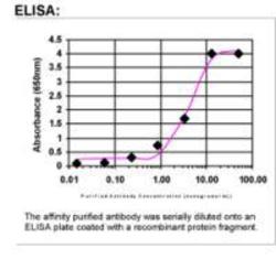
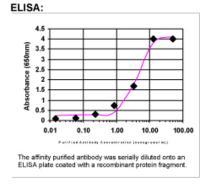
- ELISA: cleavage stimulation factor Antibody
- Validation comment
- ELISA
Supportive validation
- Submitted by
- OriGene (provider)
- Main image
- Experimental details
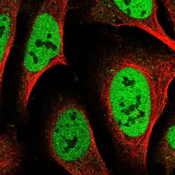
- Immunofluorescent staining of human cell line U-2 OS shows positivity in nucleus but not nucleoli.This validation was performed by Protein Atlas and the presentation of data is for informational purposes only.
- Validation comment
- IF

Supportive validation
- Submitted by
- OriGene (provider)
- Main image

- Experimental details
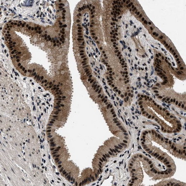
- Immunohistochemical staining of human gall bladder shows strong nuclear staining with an additional slightly weaker cytoplasmic positivity in glandular cells.This validation was performed by Protein Atlas and the presentation of data is for informational purposes only.
- Validation comment
- IHC