 Explore
 Validate
 Learn
Learn Western blot
Antibody data
- Antibody Data
- Antigen structure
- References [0]
- Comments [0]
- Validations
- Western blot [3]
- Immunoprecipitation [1]
- Immunohistochemistry [5]
Submit
Validation data
Reference
Comment
Report error
- Product number
- NB100-60443 - Provider product page

- Provider
- Novus Biologicals
- Proper citation
- Novus Cat#NB100-60443, RRID:AB_905337
- Product name
- Rabbit Polyclonal cleavage stimulation factor Antibody
- Antibody type
- Polyclonal
- Description
- Immunogen affinity purified.
- Reactivity
- Human, Mouse
- Host
- Rabbit
- Isotype
- IgG
- Vial size
- 100 ul
- Concentration
- 1.0 mg/ml
- Storage
- Store at 4C. Do not freeze.
No comments: Submit comment
Supportive validation
- Submitted by
- Novus Biologicals (provider)
- Main image
- Experimental details
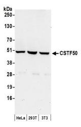
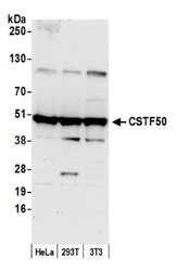
- Western Blot: cleavage stimulation factor Antibody [NB100-60443] - Detection of Human and Mouse CSTF50 by Western Blot. Samples: Whole cell lysate (50 ug) from HeLa, 293T, and mouse NIH3T3 cells prepared using NETN lysis buffer. Antibody: Affinity purified rabbit anti-CSTF50 antibody NB100-60443 used for WB at 0.4 ug/ml. Detection: Chemiluminescence with an exposure time of 30 seconds.
- Submitted by
- Novus Biologicals (provider)
- Main image
- Experimental details
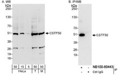
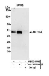
- Western Blot: cleavage stimulation factor Antibody [NB100-60443] - Whole cell lysate from HeLa (5, 15 and 50 ug for WB; 1 mg for IP, 20% of IP loaded), 293T (T; 50 ug), and mouse NIH3T3 (M; 50 ug) cells. NB100-60443 used for WB at 0.4 ug/ml (A) and 1 ug/ml (B). CSTF50 was immunoprecipitated by rabbit anti-CSTF50 antibody NB100-60444, which recognizes a downstream epitope.
- Submitted by
- Novus Biologicals (provider)
- Main image
- Experimental details
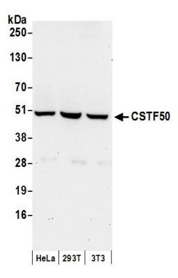
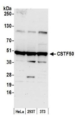
- Western Blot: cleavage stimulation factor Antibody [NB100-60443] - Detection of human and mouse CSTF50 by western blot. Samples: Whole cell lysate (50 ug) from HeLa, HEK293T, and mouse NIH 3T3 cells prepared using NETN lysis buffer. Antibody: Affinity purified rabbit anti-CSTF50 antibody NB100-60443 used for WB at 0.4 ug/ml. Detection: Chemiluminescence with an exposure time of 30 seconds.
Supportive validation
- Submitted by
- Novus Biologicals (provider)
- Main image
- Experimental details
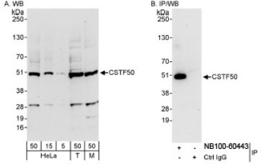
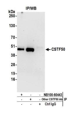
- Immunoprecipitation: cleavage stimulation factor Antibody [NB100-60443] - Detection of human CSTF50 by western blot of immunoprecipitates. Samples: Whole cell lysate (0.5 or 1.0 mg per IP reaction; 20% of IP loaded) from Hela cells prepared using NETN lysis buffer. Antibodies: Affinity purified rabbit anti-CSTF50 antibody NB100-60443 used for IP at 6 ug per reaction. CSTF50 was also immunoprecipitated by another rabbit anti-CSTF50 antibody. For blotting immunoprecipitated CSTF50, NB100-60443 was used at 1 ug/ml. Detection: Chemiluminescence with an exposure time of 30 seconds.
Supportive validation
- Submitted by
- Novus Biologicals (provider)
- Main image

- Experimental details
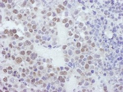
- Immunohistochemistry: cleavage stimulation factor Antibody [NB100-60443] - Sample: FFPE section of human breast carcinoma. Antibody: Affinity purified rabbit anti-CSTF50 used at a dilution of 1:1,000 (1ug/ml). Detection: DAB
- Submitted by
- Novus Biologicals (provider)
- Main image
- Experimental details
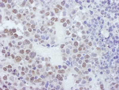
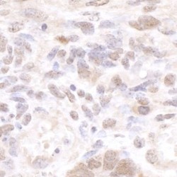
- Immunohistochemistry: cleavage stimulation factor Antibody [NB100-60443] - Sample: FFPE section of mouse plasmacytoma. Antibody: Affinity purified rabbit anti-CSTF50 used at a dilution of 1:1,000 (1ug/ml). Detection: DAB
- Submitted by
- Novus Biologicals (provider)
- Main image
- Experimental details
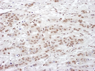
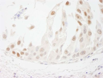
- Immunohistochemistry-Paraffin: cleavage stimulation factor Antibody [NB100-60443] - FFPE section of human ovarian carcinoma. NB100-60443 used at a dilution of 1:250.

- Submitted by
- Novus Biologicals (provider)
- Main image
- Experimental details
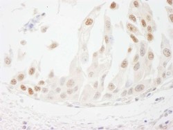
- Immunohistochemistry-Paraffin: cleavage stimulation factor Antibody [NB100-60443] - Human bladder cell carcinoma. Antibody used at a dilution of 1:1000 (1ug/ml).
- Submitted by
- Novus Biologicals (provider)
- Main image
- Experimental details
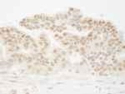
- Immunohistochemistry: cleavage stimulation factor Antibody [NB100-60443] - Detection of mouse CSTF50 by immunohistochemistry. Sample: FFPE section of mouse teratoma. Antibody: Affinity purified rabbit anti-CSTF50 (NB100-60443). Detection: DAB
