 Explore
 Validate
 Learn
Learn Western blot
 Immunohistochemistry
Antibody data
- Antibody Data
- Antigen structure
- References [1]
- Comments [0]
- Validations
- Immunohistochemistry [1]
- Flow cytometry [1]
Submit
Validation data
Reference
Comment
Report error
- Product number
- AF4398 - Provider product page

- Provider
- R&D Systems
- Product name
- Mouse CD39/ENTPD1 Antibody
- Antibody type
- Polyclonal
- Description
- Immunogen affinity purified. Detects mouse CD39/ENTPD1 in direct ELISAs and Western blots. In direct ELISAs, approximately 5% cross-reactivity with recombinant human (rh) CD39 and less than 1% cross-reactivity with rhCD39L2, rhCD39L3 and recombinant mouse CD39L3.
- Reactivity
- Mouse
- Host
- Sheep
- Conjugate
- Unconjugated
- Antigen sequence
Q921Q6- Isotype
- IgG
- Vial size
- 100 ug
- Concentration
- LYOPH
- Storage
- Use a manual defrost freezer and avoid repeated freeze-thaw cycles. 12 months from date of receipt, -20 to -70 °C as supplied. 1 month, 2 to 8 °C under sterile conditions after reconstitution. 6 months, -20 to -70 °C under sterile conditions after reconstitution.
Submitted references Complete deletion of Cd39 is atheroprotective in apolipoprotein E-deficient mice.
De Giorgi M, Enjyoji K, Jiang G, Csizmadia E, Mitsuhashi S, Gumina RJ, Smolenski RT, Robson SC
Journal of lipid research 2017 Jul;58(7):1292-1305
Journal of lipid research 2017 Jul;58(7):1292-1305
No comments: Submit comment
Supportive validation
- Submitted by
- R&D Systems (provider)
- Main image
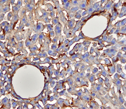
- Experimental details
- CD39/ENTPD1 in Mouse Liver. CD39/ENTPD1 was detected in perfusion fixed frozen sections of mouse liver using Mouse CD39/ENTPD1 Antigen Affinity-purified Polyclonal Antibody (Catalog # AF4398) at 15 µg/mL overnight at 4 °C. Tissue was stained using the Anti-Sheep HRP-DAB Cell & Tissue Staining Kit (brown; Catalog # CTS019) and counterstained with hematoxylin (blue). View our protocol for Chromogenic IHC Staining of Frozen Tissue Sections.

Supportive validation
- Submitted by
- R&D Systems (provider)
- Main image
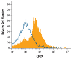
- Experimental details
- Detection of CD39/ENTPD1 in Mouse Splenocytes by Flow Cytometry. Mouse splenocytes were stained with Mouse CD39/ENTPD1 Antigen Affinity-purified Polyclonal Antibody (Catalog # AF4398, filled histogram) or control antibody (Catalog # 5-001-A, open histogram), followed by NorthernLights™ 637-conjugated Anti-Sheep IgG Secondary Antibody (Catalog # NL011).
